. 2008 Nov;39(3):342-55.
doi: 10.1016/j.mcn.2008.07.014. Epub 2008 Jul 29.

Deletion of lysophosphatidic acid receptor LPA1 reduces neurogenesis in the mouse dentate gyrus

Affiliations

Deletion of lysophosphatidic acid receptor LPA1 reduces neurogenesis in the mouse dentate gyrus

Elisa Matas-Rico et al. Mol Cell Neurosci. 2008 Nov.

Abstract

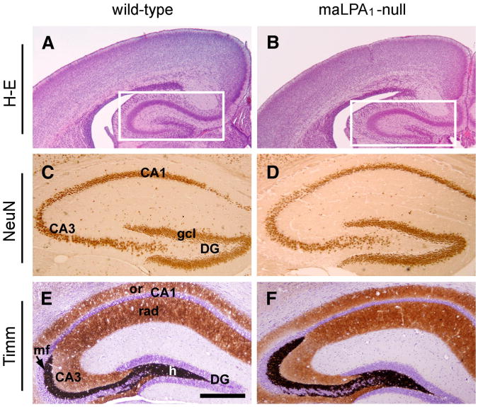
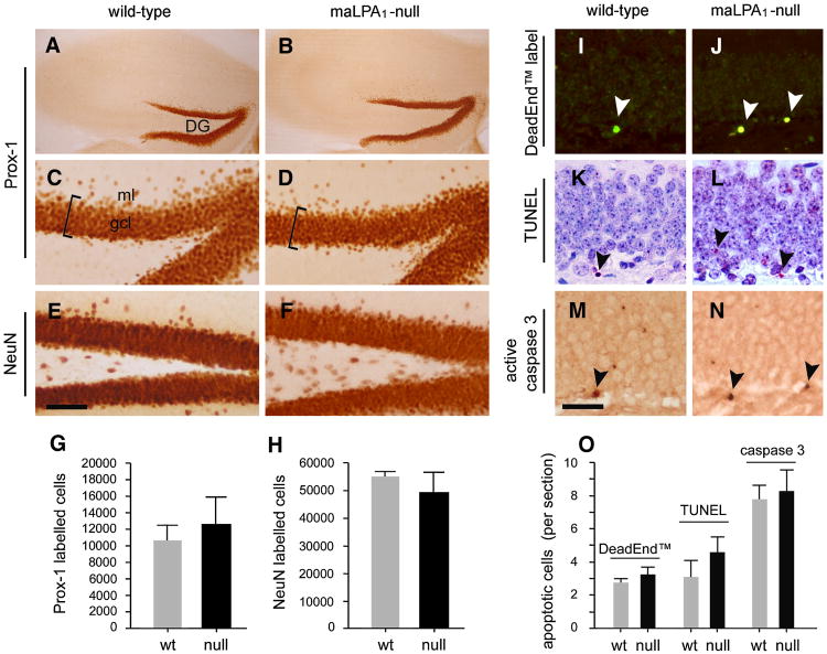
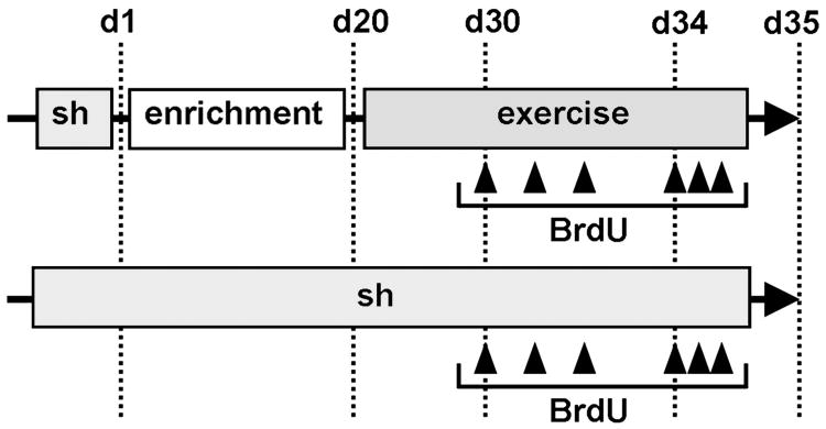
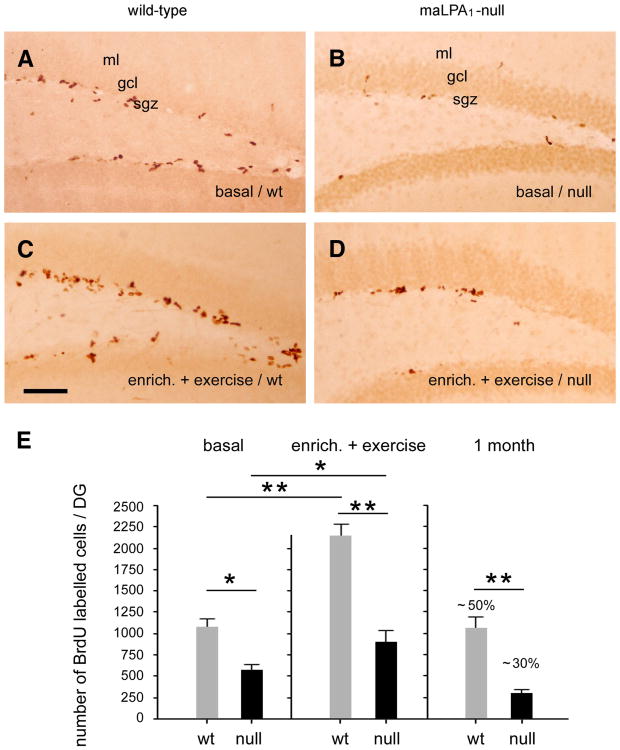
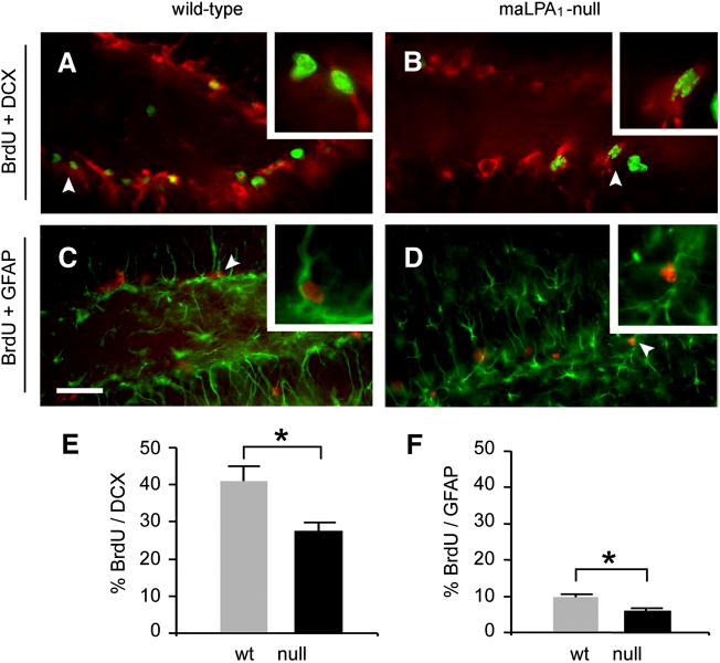
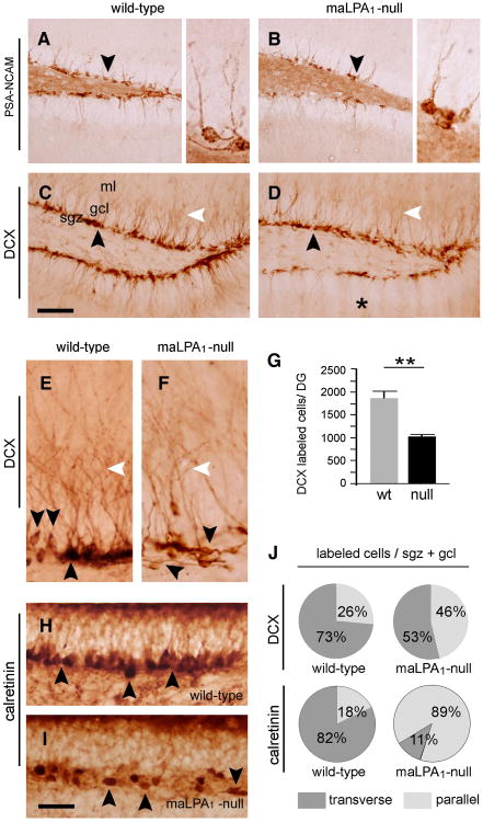
Neurogenesis persists in certain regions of the adult brain including the subgranular zone of the hippocampal dentate gyrus wherein its regulation is essential, particularly in relation to learning, stress and modulation of mood. Lysophosphatidic acid (LPA) is an extracellular signaling phospholipid with important neural regulatory properties mediated by specific G protein-coupled receptors, LPA(1-5). LPA(1) is highly expressed in the developing neurogenic ventricular zone wherein it is required for normal embryonic neurogenesis, and, by extension may play a role in adult neurogenesis as well. By means of the analyses of a variant of the original LPA(1)-null mutant mouse, termed the Malaga variant or "maLPA(1)-null," which has recently been reported to have defective neurogenesis within the embryonic cerebral cortex, we report here a role for LPA(1) in adult hippocampal neurogenesis. Proliferation, differentiation and survival of newly formed neurons are defective in the absence of LPA(1) under normal conditions and following exposure to enriched environment and voluntary exercise. Furthermore, analysis of trophic factors in maLPA(1)-null mice demonstrated alterations in brain-derived neurotrophic factor and insulin growth factor 1 levels after enrichment and exercise. Morphological analyses of doublecortin positive cells revealed the anomalous prevalence of bipolar cells in the subgranular zone, supporting the operation of LPA(1) signaling pathways in normal proliferation, maturation and differentiation of neuronal precursors.


Figures

Fig.1
Fig.1. Training schedule and BrdU administration
Schematic representation of the experimental design (see Materials and methods for details). BrdU was i.p. injected (arrowheads) during four days (days 30-34) after a period of exposure to environmental enrichment (d1 to d20) followed by voluntary exercise (days 20-30). Exercise was maintained during days of injection. Naïve animals were under standardized housing (Sh) and receive appropriate BrdU doses likewise on an equivalent period. On day 35 animals were processed for fixation.
Fig.2
Fig.2. Hippocampus formation in wild-type and maLPA1-null mice
Comparable hippocampal DG coronal sections from wild-type (A, C, E) and maLPA1-null (B, D, F) mice at postnatal P0 (A, B) and 12 weeks-old (E-F) ages. (A, B) Haematoxylin-stained wild-type (A) and maLPA1-null (B) cerebral cortices. Hippocampal formation is marked in rectangles. (C, D) Neuronal expression of NeuN in postnatal P0 mice states clearly and evidences a normal granule cell layer (gcl) in dentate gyrus (DG), CA3 and CA1 hippocampal areas in wild-type (C) and maLPA1 null (D) mice. (E, F) Timm's stained sections from wild-type (E) and maLPA1-null (F) mice. Reactive zinc labels the hilus (h), the mossy fibers (mf) into CA3 area and shows a lighter laminar staining in stratum radiatum (rad) and oriens (or) of CA1. Both genotypes exhibit a similar histochemical pattern. Scale bar in A, B = 1000 μm; C, D = 375 μm; E, F = 300 μm.
Fig.3
Fig.3. Normal development and apoptosis of mature granule cell layer of maLPA1-null mice
(A, B, C, D) Immunolabelling of granule cells with the specific marker Prox-1 in 3 months-old mice reveals no differences in coronal sections of the DG from maLPA1-null hippocampus (B, detailed in D) when compared with wild-type (A, detailed in C). gcl, granule cell layer, bracket delimits layer thickness; ml, molecular layer. (E, F) Similar results were obtained after immunostaining in parallel sections with the neuronal marker NeuN in wild-type (E) or maLPA1-null mice (F). (G, H) Total number of Prox-1 positive (G) or NeuN positive (H) granule cells in DG of wild-type (wt) and maLPA1-null (null) mice. Analysis did not found significant differences between both genotypes (n = 6). (I-O) Cell death detection in wild-type and maLPA1-null mice. Representative photographs of 3 months-old mice coronal hippocampal sections from wild-type (I, K, M) and maLPA1-null (J, L, N) mice showing labelled nuclei (arrowheads, as examples) with DeadEnd™ Colorimetric Apoptosis Detection System (I, J), TUNEL (K, L) or anti-active caspase 3 (M, N). (O) Cell death detection in wild-type and maLPA1-null DG expressed as number of apoptotic cells per section for each corresponding method. LPA1-null DG exhibits a scarcely detectable increased levels of apoptosis although without statistical variation from observed in wild-type (n= 8). In both graphs data expressed as mean ± SEM. Scale bar in A, B = 540 μm; C-F = 90 μm; I-N = 270 μm.
Fig.4
Fig.4. Reduced cell proliferation and production of neural cells in the dentate gyrus in absence of LPA1
(A-D) Representative immunohistochemical staining for BrdU detection in adult hippocampus. The basal number of BrdU-positive cells (A) is increased in the wild-type mice after exposure at enriched environment for 20 days followed by 14 days of activity in the running-wheel (C). By contrast, basal neural precursor proliferation is reduced in the DG of maLPA1-null mice (B) in almost 50% from the total label as compared with the wild-type. This reduction is maintained even in housing conditions of environmental enrichment and exercise (D). (E) Estimation of numbers of total BrdU labelled cells in wild-type (wt) and maLPA1-null (null) adult mice for i) basal condition (basal), ii) after the exposure at enriched environment and exercise (enrich+ exercise), and iii) one-month later of last BrdU injection, to evaluate the survival of generated cells. Bars represent mean ± SEM. Reduction of cell proliferation was significant between both genotypes in all conditions (n= 8; *, P< 0.05; **, P < 0.01). Significantly, whilst wild-type mice increase the number of BrdU positive cells under environmental complexity and exercise, (n= 8; **, P < 0.05) maLPA1-null mice do not initiate a significant proliferation under these conditions. Likewise, survival of newly generated cells is clearly minor in absence of LPA1 receptor, with values estimated in about 20% less than wild-type (n= 8; *, P< 0.01). Abbreviations as in previous figures. Scale bar = 100 μm.
Fig.5
Fig.5. Phenotypic characterization of newly generated dentate gyrus cells
(A, B) Descriptive images of double immunofluorescence labelling for newly born DG cells (BrdU positive, Alexafluor® 488) with early neuronal phenotype (doublecortin, DCX positive, Alexafluor® 568) in wild-type (A) and maLPA1-null (B) mice after exposure to environmental enrichment and exercise. (C, D) Equivalent DG sections double immunolabelled for newly born cells (BrdU positive, Alexafluor® 568) with glial phenotype (GFAP positive, Alexafluor® 488) in wild-type (C) and maLPA1-null (D) mice. Arrowheads magnified in squares. (E, F) Analysis of the percentages of double labelled cells (expressed as mean ± SEM, n= 10; * P < 0.05) shows similar neuronal/glial proportions in newly born DG cells from wild-type and maLPA1-null mice. Data for neuronal (DCX positive, E) or glial (GFAP positive, F) phenotype show equally preferential neuronal fate against glial although the percentages differ significantly between both genotypes with less amount of double DCX or GFAP BrdU labelled cells in absence of LPA1. Scale bar = 200 μm; small images in A-D, 3× magnified.
Fig.6
Fig.6. Reduced PSA-NCAM and doublecortin expression and altered expression pattern in absence of LPA1receptor
(A, B) Illustrations of PSA-NCAM labelled cells in the dentate gyrus of wild-type (A) and maLPA1-null (B) adult mice following enrichment and voluntary exercise. Expression of PSA-NCAM by newly born cells correlates with the reduced proliferation in mice lacking LPA1 receptor showing less number of stained cells (arrowheads). Adjacent images in A and B are 6× detailed from arrowhead-pointed. PSA-NCAM immunolocalization delimits the cell body and extending process in both genotypes. (C-F) Photomicrographs illustrates the doublecortin expression and the morphology of early generated DG neurons of wild-type (C, E) and maLPA1-null (D, F) adult mice in same conditions. A lesser amount of DCX positive cells (black arrowheads) and dendritic processes extending into the molecular layer through GCL (white arrowheads) are observed in maLPA1-null mice, being particularly reduced in the lower blade of hippocampus (asterisk). Enlarged images of SGZ/GCL (E, F) show a reduced cellular branching in maLPA1-null DG as well as a preference of bipolar tangential cells with cell processes running parallel to SGZ/GCL (black arrowheads) against radially oriented that predominate in same wild-type area (black arrowheads). (G) Estimation of numbers of total dentate DCX labelled cells in wild-type (wt) and maLPA1-null (null) adult mice (n= 8; **, P < 0.01). (H, I) Calretinin expression at SGZ/GCL of wild-type (H) and maLPA1-null (I) mice defines the different disposition of cell processes. Arrowheads point the soma of immunoreactive cells. (J) Estimation of percentages of SGZ/GCL DCX and calretinin labelled cells in wild-type (wt) and maLPA1-null (null) adult mice showing transverse (radial) or parallel (tangential) to GCL cell processes (n= 10; significance was found among wild-type and maLPA1-null mice, P < 0.05). gcl, granule cell layer; ml, molecular layer; sgz, subgranular zone. Scale bar in A, B = 200 μm; C, D = 175 μm; E, F, H, I = 35 μm.
Fig.7
Fig.7. Vascularity and analysis of the trophic factors in the maLPA1-null hippocampus
(A, B) Double immunodetection for BrdU positive proliferating cells (revealed with DAB plus nickel; black-purple reaction) and lectin histochemistry for blood vessels (DAB; brown reaction) in the dentate gyrus of wild-type (A) and maLPA1-null (B) mice. Subgranular zone (sgz) proliferating cells (arrowheads) mostly associate with vessels in both genotypes as seen in corresponding magnified image at left squares. (C) Graph shows the quantified percentage of subgranular cells proliferating cells associated to vessels, expressed as mean ± SEM (n=10). No significant differences were found in absence of LPA1 receptor (null) with almost 80% of cells adjacent to vascular in the same way to wild-type hippocampus (wt). (D) BDNF protein levels in hippocampus measured by ELISA for wild-type (wt) and maLPA1-null (null) mice in basal and following enrichment and voluntary exercise. Data, indicated as mean ± SEM, do not differ between both groups at basal condition showing normal BDNF levels in maLPA1-null mice as compared with control. Exposure to enriched environment and voluntary exercise raised the protein levels in both wild-type and maLPA1-null mice, though this increase was significantly superior in wild-type mice (n= 8; * P < 0.05;** P < 0.001). (D) NGF protein levels in hippocampus measured by ELISA for wild-type (wt) and maLPA1-null (null) mice in basal and following enrichment and voluntary exercise. Data, indicated as mean ± SEM, show the differences between both groups at basal condition and experimental conditions. Exposure to enriched environment and voluntary exercise made significant the increase of NGF levels in absence of LPA1. (n= 8; * P < 0.05). (E) Likewise, the enzimoimmunoassay for IGF-I protein level determination in similar extracts showed increase of IGF-I and differences between both genotypes just after experience and exercise. Remarkably, the increase of IGF-I levels in maLPA1-null protein samples doubled the observed for wild-type samples (n= 8; ** P < 0.01). Scale bar in A, B = 135 μm (small images 4× augmented).

Similar articles

Cited by

References

    1. Åberg MAI, Åberg ND, Palmer TD, Alborn A, Carlsson-Skwirut C, Bang P, Rosengren LE, Olsson T, Gage FH, Eriksson PS. IGF-I has a direct proliferative effect in adult hippocampal progenitor cells. Mol Cell Neurosci. 2003;24:2–40. - PubMed
    1. Abrous DN, Koehl M, Le Moal M. Adult neurogenesis: from precursors to network and physiology. Physiol Rev. 2005;85:523–569. - PubMed
    1. Ahn S, Joyner AL. In vivo analysis of quiescent adult neural stem cells responding to Sonic hedgehog. Nature. 2005;437:894–897. - PubMed
    1. Aimone JB, Wiles J, Gage FH. Potential role for adult neurogenesis in the encoding of time in new memories. Nat Neurosci. 2006;9:723–727. - PubMed
    1. Allard J, Barrón S, Diaz J, Lubetzki C, Zalc B, Schwartz JC, Sokoloff P. A rat G protein-coupled receptor selectively expressed in myelin-forming cells. E J Neurosci. 1998;10:1045–1053. - PubMed

Publication types

Substances