. 2008 Nov;9(11):1036-48.
doi: 10.1016/j.jpain.2008.06.006. Epub 2008 Aug 16.

Partial infraorbital nerve ligation as a model of trigeminal nerve injury in the mouse: behavioral, neural, and glial reactions

Affiliations

Partial infraorbital nerve ligation as a model of trigeminal nerve injury in the mouse: behavioral, neural, and glial reactions

Mei Xu et al. J Pain. 2008 Nov.

Abstract

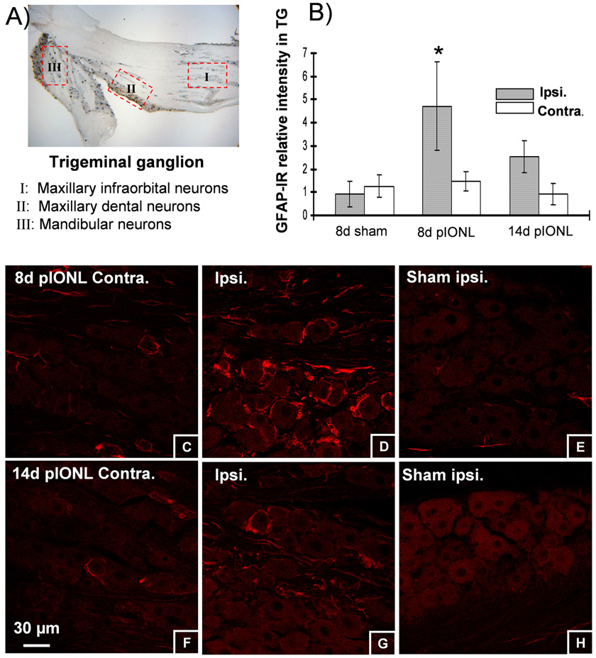
Trigeminal nerve damage often leads to chronic pain syndromes including trigeminal neuralgia, a severely debilitating chronic orofacial pain syndrome. Options for treatment of neuropathic pain are limited in effectiveness and new approaches based on a better understanding of the underlying pathologies are required. Partial ligation has been shown to effectively mimic many of the qualities of human neuropathic pain syndromes. We have devised a mouse model of trigeminal neuralgia using a partial infraorbital nerve ligation (pIONL) that induces persistent pain behaviors and morphological changes in the brainstem. We found that the pIONL effectively induced mechanical allodynia lasting for more than 3 weeks. Cell proliferation (bromodeoxyuridine), activation of astrocytes and microglia in the ipsilateral caudal medulla, and persistent satellite cell reaction in the ipsilateral ganglion were observed. Neurochemical markers calcitonin gene-related peptide, substance P were decreased in medullary dorsal horn ipsilateral to the injury side, whereas substance P receptor NK1 expression was increased after 8 days. Nerve injury marker ATF3 was markedly increased in ipsilateral trigeminal ganglion neurons at 8 days after pIONL. The data indicate that partial trigeminal injury in mice produces many persistent anatomical changes in neuropathic pain, as well as mechanical allodynia.

Perspective: This study describes the development of a new mouse model of trigeminal neuropathic pain. Our goal is to devise better treatments of trigeminal pain, and this will be facilitated by characterization of the underlying cellular and molecular neuropathological mechanisms in genetically designed mice.


Figures

Fig 1
Fig 1. Images showing the surgical approach used during partial infraorbital nerve ligation (pIONL) and mechanical allodynia produced following pIONL
(A) The exposure of right ION (arrow) and tight ligature with 7-0 thread (arrowhead) at the lateral half of nerve. Scale bar: 2 mm. (B) Response thresholds to usually innocuous tactile (von Frey hair) stimuli in sham and pIONL mice during the 25 days after surgery. Mice with pIONL developed significant allodynia as evident from the decreased thresholds to tactile stimulation compared with mice receiving sham surgeries. The mechanical allodynia for ligated mice lasted for >3 weeks. Asterisks indicate significant decreases in response thresholds in pIONL-operated mice compared to the sham-operated mice on the days noted (*P<0.001, ANOVA, non-parametric analysis). Data are presented as the mean±SEM von Frey hair threshold in grams; n=6–8 per time point.
Fig 2
Fig 2. Change in the duration of face grooming behaviors and body weight after pIONL
(A) The total time for isolated face grooming episodes during a 15 min observation period was elevated for the pIONL mice only during the first day after surgery. (*P<0.05, ANOVA followed by Student-Newman–Keuls test, n=4 per group). (B) Sham operated mice were gaining weight by 3 days after surgery, whereas pIONL mice lost less than 5% during the first week and then started to gain weight during the second week. (n=6–8 per group)
Fig 3
Fig 3. Neuronal expression of CGRP-IR, sub P-IR and NK1R-IR in the cervical C1 spinal cord and caudal medulla at 8 days after pIONL
There was a decrease of CGRP-IR (A, B), sub P-IR (E, F, G) and increase of NK1 receptor-IR (C, D) in the superficial dorsal horn of C1 spinal cord and caudal medulla ipsilateral to the nerve injury. A, C are cervical C1 spinal cord sections; B, D, E are caudal medulla sections. F, G are the high magnifications of the dorsal horn in E. H, I, J graphs showing mean ± SEM pixel intensity of CGRP, NK1-R and Sub P staining in caudal medulla lamina brainstem sections. H, image analysis of CGRP expression in ipsilateral caudal medulla after pIONL demonstrated approximately 1.5 fold lower signal intensity [2.17 ± 0.13 arbitrary units (AU); n = 20 sections from 4 animals] than that of contralateral caudal medulla (3.27 ± 0.11 AU; n = 20 sections from 4 animals, *p < 0.001) or sham-ligated mice (3.16 ± 0.12 AU; n = 20 sections from 4 animals, **p < 0.001). I, image analysis of NK1-receptor staining in ipsilateral caudal medulla after pIONL demonstrated approximately 1.26 fold higher signal intensity (1.662 ± 0.06 AU; n = 20 sections from 4 animals) than that of contralateral caudal medulla (1.309 ± 0.1AU; n = 20 sections from 4 animals, *p < 0.001) or sham-ligated mice (1.33 ± 0.17 AU; n = 20 sections from 4 animals, **p < 0.001). J, image analysis of Sub P expression in ipsilateral caudal medulla after pIONL demonstrated approximately 1.3 fold lower signal intensity (1.50 ± 0.04 AU; n = 20 sections from 4 animals) than that of contralateral caudal medulla (1.98 ± 0.07AU; n = 20 sections from 4 animals, *p < 0.001) or sham-ligated mice (1.99 ± 0.16 AU; n = 20 sections from 4 animals, **p < 0.001). Scale bars: 400 µm
Fig 4
Fig 4. Microglial responses after pIONL
(A) pIONL induced microglial activation (CD11b staining) in the ipsilateral side of the caudal medulla. A representative, higher power image of a microglial cell is shown (B). Higher magnification images (taken from the boxes outlined in panel A), show differences in contralateral (C) and ipsilateral (D) CD11b-IR 1d after pIONL. In contrast, CD11b-IR in the caudal medulla was not different in the contralateral (E) or ipsilateral (E) 8d after pIONL The microglial activation had decreased by 8d after surgery (E, F). Quantification showing mean ± SEM pixel intensity of CD11b staining in caudal medulla of brainstem sections (G). Image analysis of CD11b expression in ipsilateral caudal medulla 1 day after pIONL demonstrated approximately 1.25 fold higher signal intensity (1.67 ± 0.07 AU; n = 16 sections from 4 animals) than that of contralateral caudal medulla (1.32 ± 0.04AU; n = 16 sections from 4 animals, *p < 0.001) or sham-ligated mice (1.24 ± 0.03 AU; n = 16 sections from 4 animals, **p < 0.001). In contrast, image analysis of CD 11b staining in caudal medulla 8 days after pIONL did not show a significant increase over sham mice (p > 0.05). Scale bars: A, 400 µm; B, 15 µm; C–F, 30 µm.
Fig 5
Fig 5
Astrocytic responses in caudal medulla 8d after pIONL (A). GFAP staining was compared in ipsi and contraleteral regions of the caudal medulla of sham operated mice (B, C) and following pIONL (D, E). There was an striking increase of GFAP-IR on the ipsilateral side of caudal medulla (E) compared with contralateral side (D). The response involved hypertrophy of the ipsilateral astrocytes (E). Image analysis of GFAP expression in ipsilateral caudal medulla 8 day after pIONL (F) demonstrated approximately 2 fold higher signal intensity (8.37 ± 0.43 AU; n = 20 sections from 4 animals) than that of contralateral caudal medulla (4.78 ± 0.66 AU; n = 20 sections from 4 animals, *p < 0.001) or sham-ligated mice (3.88 ± 0.86 AU; n = 20 sections from 4 animals, **p < 0.001). Scale bars: A, 400 µm; B–E 30 µm.
Fig 6
Fig 6. Cellular proliferation in caudal medulla 8d after pIONL was detected following daily BrdU administration (100 mg/kg intraperitoneally, once daily for 8 days)
A, BrdU-positive cells were markedly increased on the ipsilateral side of the caudal medulla compared with the contralateral side. B, a summary graph showing mean ± SEM BrdU positive cells in caudal medulla of brainstem sections. Image analysis of BruU positive cells in ipsilateral caudal medulla 8 day after pIONL demonstrated approximately 7 fold higher BrdU-IR (34.75 ± 8.4; n = 8 sections from 4 animals) than that of the contralateral caudal medulla (6.12 ± 1.4; n = 8 sections from 4 animals, *p < 0.001) or sham-ligated mice (5 ± 1.8; n = 8 sections from 4 animals, **p < 0.001). C–E, Some of the dividing BrdU-labeled cells were also CD 11b positive microglia (arrows), and others were not CD 11b positive (arrowheads). F–H, Some of the dividing BrdU-labeled cells were GFAP-positive astrocytes (arrows), and others were not GFAP positive (arrowheads). I–K, Many BrdU-positive cells were obviously double labeled with nestin, a stem cell marker (arrows), but a small fraction of BrdU-positive cells were not nestin-positive stem cells (arrowheads). L–N, BrdU-labeled cells were not co-labeled by NG2, an oligodendrocyte precursor marker (L–N, arrowheads). Scale bars: A, 200 µm; C–N 100 µm.
Fig 7
Fig 7. GFAP positive satellite cell changes in trigeminal ganglion 1wk and 2wks after pIONL
Upper left panel: Neuronal cell body distribution in trigeminal ganglion. I: Maxillary infraorbital neurons. II: Maxillary dental neurons. III: Mandibular neurons. Our quantitation was for zone (I) for 3 sections (20 µm thickness) per ganglion, with intervals of 60 µm between each counted section. Upper right panel: Quantification of satellite cell reaction in zone (I) of TG, *p<0.05, n=4. Lower panel: GFAP-IR in the trigeminal ganglion of wild-type mouse 1 wk and 2 wks after pIONL. There is an apparent increase in GFAP-IR in the ipsilateral side of TG (D) compared with the contralateral side (C) or sham mice (E) 1wk after surgery. The GFAP-IR decreases 2 wks after pIONL (F, G). Scale bars, C–H, 30 µm.
Fig 8
Fig 8. Immunohistochemical expression of ATF3 in trigeminal ganglia 8 days after pIONL
ATF3-IR increased markedly in the ipsilateral TG (B,D) compared with the contralateral TG(A,C). ATF3 expression occurred within neurons in the infraorbital region of TG (B). E, summary graph shows quantification (mean ± SEM) of ATF3 positive cells in caudal medulla of brainstem sections. Image analysis of ATF3 positive cells in ipsilateral caudal medulla 8 days after pIONL demonstrated approximately 10 fold higher (52 ± 6.64; n = 8 sections from 4 animals) than that in contralateral caudal medulla (5.8 ± 1.59; n = 8 sections from 4 animals, *p < 0.001) or shamligated mice (3.9± 0.69; n = 8 sections from 4 animals, **p < 0.001). Scale bars: A,B, 500 µm; C,D, 200 µm.

References

    1. Abbadie C, Brown JL, Mantyh PW, Basbaum AI. Spinal cord substance P receptor immunoreactivity increases in both inflammatory and nerve injumry models of persistent pain. Neuroscience. 1996;70:201–209. - PubMed
    1. Anderson LC, von Bartheld CS, Byers MR. NGF depletion reduces ipsilateral and contrlateral trigeminal satellite cell reactions after inferior alveolar nerve injury in adult rats. Exp Neurol. 1998;150:312–320. - PubMed
    1. Anderson LC, Vakoula A, Veinote R. Inflammatory hypersensitivity in a rat model of trigeminal neuropathic pain. Arch Oral Biol. 2003;48:161–169. - PubMed
    1. Averill S, Michael GJ, Shortland PJ, Leavesley RC, King VR, Bradbury EJ, McMahon SB, Priestley JV. NGF and GDNF ameliorate the increase in ATF3 expression which occurs in dorsal rootganglion cells in response to peripheral nerve injury. Eur. J. Neurosci. 2004;19:1437–1445. - PubMed
    1. Bennett GJ, Xie YK. A peripheral mononeuropathy in rat that produces disorders of pain sensation like those seen in man. Pain. 1988;33:87–107. - PubMed

Publication types

MeSH terms