Review
. 2008 Dec 6;5(29):1391-408.
doi: 10.1098/rsif.2008.0185.

Development of free-energy-based models for chaperonin containing TCP-1 mediated folding of actin

Affiliations
Review

Development of free-energy-based models for chaperonin containing TCP-1 mediated folding of actin

Gabriel M Altschuler et al. J R Soc Interface. .

Abstract

A free-energy-based approach is used to describe the mechanism through which chaperonin-containing TCP-1 (CCT) folds the filament-forming cytoskeletal protein actin, which is one of its primary substrates. The experimental observations on the actin folding and unfolding pathways are collated and then re-examined from this perspective, allowing us to determine the position of the CCT intervention on the actin free-energy folding landscape. The essential role for CCT in actin folding is to provide a free-energy contribution from its ATP cycle, which drives actin to fold from a stable, trapped intermediate I3, to a less stable but now productive folding intermediate I2. We develop two hypothetical mechanisms for actin folding founded upon concepts established for the bacterial type I chaperonin GroEL and extend them to the much more complex CCT system of eukaryotes. A new model is presented in which CCT facilitates free-energy transfer through direct coupling of the nucleotide hydrolysis cycle to the phases of actin substrate maturation.


Figures

Figure 1
Figure 1
Chaperonin architecture. Schematic demonstrating the quaternary structure of type I and type II chaperonins based on their respective atomic structures (Braig et al. 1994; Ditzel et al. 1998). (a) The type I chaperonin GroEL consists of two rings of seven identical subunits. Inter-ring contacts occur via the equatorial domains, with each subunit interacting with two subunits in the opposite ring. Conformational changes relating to the identity of the nucleotide bound within the equatorial domain are transmitted via the intermediate domain to the apical domain. The apical domain interacts with the co-chaperonin GroES, consisting of seven identical subunits, which caps one end of the chaperonin during a folding cycle. (b) The thermosome is a type II chaperonin consisting of two rings of eight subunits that alternate between α and β isoforms (cylinder dimensions 16×15 nm). The distribution of domains within each subunit is the same as that for GroEL; however, each subunit interacts with only one subunit in the opposite ring. There is no associated co-chaperonin; however, the apical domains can adopt a conformation that effectively caps the end of the chaperonin in a similar manner to that of GroES (not shown in figure).
Figure 2
Figure 2
Free-energy representation of GroEL folding models. (a) One-dimensional free-energy surface of a hypothetical chaperonin substrate demonstrating relative positions of the unfolded state U, a kinetically trapped folding intermediate IT, a productive folding intermediate with higher free energy IP, and the native state N, the global energy minimum. Autonomous folding of the substrate from U proceeds to the local minimum IT. A high activation energy barrier partitions this state from further folding intermediates. (b) The kinetic model; the chaperonin acts as a catalyst. Substrate binding reduces the kinetic energy barrier (solid line) between the intermediates IT and IP relative to the original energy surface (dashed line), allowing significant population of the IP conformation, from which folding to N occurs—either bound or not bound to the chaperonin walls. (c) The thermodynamic model; binding to GroEL modifies the energy landscape (solid line) and stabilizes the intermediate IP relative to IT and N, resulting in population of IP. Substrate is then released from the chaperonin walls and folding occurs from this productive intermediate on the original chaperonin free landscape (dashed line), increasing the probability of native state formation. (d) The forced unfolding model; initially, binding of substrate to chaperone is favoured due to the low free energy of the chaperonin–substrate complex (red line). Once bound, ATP-powered conformational rearrangements of the chaperone directly drive modification of the substrate energy levels to a new energy landscape (arrow to black solid line), resulting in ‘unfolding’ from IT to IP. In a similar manner to the kinetic and thermodynamic examples (b and c), release of IP from the chaperonin walls results in spontaneous folding to the native state on the chaperonin-free landscape.
Figure 3
Figure 3
Structures of actin in folded and CCT-bound conformations. (a) Atomic structure of rabbit skeletal muscle G-actin taken from Kabsch et al. (1990; PDB code 1ATN) in the standard front view with the subdomains 1–4 numbered at the corners. Ca2+ (green sphere) and nucleotide (ball and stick) are bound in the cleft between the subdomains 3 and 4 (the large domain), and 1 and 2 (the small domain). CCT-binding sites, as identified by a β-actin peptide array and site-directed mutagenesis (Hynes & Willison 2000; McCormack et al. 2001b), are coloured; site I (red), II (green) and III (cyan). The C-terminus (gold) and the highly conserved ‘hinge’ residues G146 and G150 (purple) have been highlighted. (b) Electron microscopy reconstructions of the actin–CCT complex in the (i) absence and (ii) presence of ATP (AMPPNP), demonstrating the structural changes undergone by actin during the CCT functional cycle. The atomic structure of actin has been docked to the intra-cavity volume. The N-terminal domain (red) binds to CCT with lower affinity than the C-terminal domain (white). ATP binding to CCT induces large movements of the apical domains that give rise to a more native conformation for the bound actin. This image is reproduced from Llorca et al. (2001).
Figure 4
Figure 4
Proposed one-dimensional free-energy landscape for actin folding. This representation is a one-dimensional simplification of the rough multidimensional free-energy landscape for actin. States in rapid equilibrium are grouped and labelled according to the specific folding intermediates that have been experimentally identified: I1, I2 and I3 (Altschuler et al. 2005). Monomeric, chemically unfolded or nascent actin, U/ActNasc, spontaneously folds to form the intermediate I3. At moderate to high protein concentration this species irreversibly forms aggregates, which may represent the lowest free-energy actin species. At low protein concentration, I3 is kinetically and thermodynamically isolated with respect to ligand-free folded actin I2, and further folding of actin independent of CCT is not observed on any physiologically significant time scale. The forward folding reaction (I3→I2) is associated with ATP-dependent CCT interaction and activity (arrow in blue shaded area). I2 is thermodynamically unstable with respect to the unfolded, CCT-binding, intermediate I3. Unfolding from I2 to I3 is slow at room temperature (ku∼0.01 s) due to the large activation energy of unfolding, measured as approximately 80 kJ mol−1 (Schuler et al. 2000; Altschuler et al. 2005). Once formed, I2 does not bind CCT (Rohman 1999) and spontaneously binds nucleotide and cation, stabilizing actin in a conformational ensemble referred to as the ‘native platform’ (green shaded area).
Figure 5
Figure 5
CCT free-energy cycle for a single ring. One-dimensional free-energy landscape of a four-state CCT folding cycle. The energy levels of the four CCT intermediates i–iv, and the transition states between them depend on the identity of the nucleotide bound to CCT: solid line, ATP bound; dashed line, ADP bound. When the chaperone is fully ATP bound, state iii has the lowest free energy (circled) and when fully ADP bound, state i has the lowest energy (dotted circled). The net free-energy change of CCT is zero through the CCTADP→CCTATP→CCTADP cycle. However, the topography of the ATP/ADP free-energy surfaces dictates a kinetically favoured path i→ii→iii→iv→i.
Figure 6
Figure 6
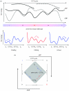
‘Active CCT’ model of actin folding: CCT couples ATP binding and hydrolysis to the CCT-associated actin free-energy landscape. (a) Strong, specific interactions between actin and CCT directly couple the actin free-energy landscape to that of the CCT cycle. As CCT binds and hydrolyses nucleotide, undergoing the conformational cycle described in figure 6, the free-energy landscape of bound actin changes depending on the CCT conformation. Here, two hypothetical actin energy landscapes are described; corresponding to actin bound to CCT intermediate i (blue) and to CCT intermediate iii (red). The free energy required to modify the actin landscape between the blue and red topographies is derived from the energetically favourable, downhill phases in the CCT cycle; i→ii→iii for CCT–ATP and iii→iv→i for CCT–ADP. The blue landscape favours binding of unfolded actin I3 and release of the folded intermediate I2 while the red landscape favours folding CCT-I3→CCT-I2. The concept of alternate landscapes proposed here is similar to that of the GroEL thermodynamic model (figure 2c). (b) The consequences of the hypothetical landscapes can be summarized in a reaction scheme within the context of the CCT–ATP/ADP cycle. The entrance and exit (binding and release) points for actin within the cycle are displayed. This scheme is cyclical from the CCT point of view but linear with respect to actin, i.e. at the end of a single cycle, the CCT regains its initial state but actin has progressed along its folding coordinate.

References

    1. Altschuler, G. M. 2006 Protein folding studies on the actin–CCT chaperone system. PhD thesis, Institute of Cancer Research, University of London.
    1. Altschuler G.M, Klug D.R, Willison K.R. Unfolding energetics of G-alpha-actin: a discrete intermediate can be re-folded to the native state by CCT. J. Mol. Biol. 2005;353:385–396. doi: 10.1016/j.jmb.2005.07.062. - DOI - PubMed
    1. Anfinsen C.B. Principles that govern the folding of protein chains. Science. 1973;181:223–230. doi: 10.1126/science.181.4096.223. - DOI - PubMed
    1. Archibald J.M, O'Kelly C.J, Doolittle W.F. The chaperonin genes of jakobid and jakobid-like flagellates: implications for eukaryotic evolution. Mol. Biol. Evol. 2002;19:422–431. - PubMed
    1. Astumian R.D. Thermodynamics and kinetics of a Brownian motor. Science. 1997;276:917–922. doi: 10.1126/science.276.5314.917. - DOI - PubMed

Publication types