. 2008 Dec 12;283(50):34469-89.
doi: 10.1074/jbc.X800009200. Epub 2008 Aug 15.

Discovery of cellular regulation by protein degradation

Affiliations

Discovery of cellular regulation by protein degradation

Alexander Varshavsky. J Biol Chem. .
No abstract available


Figures

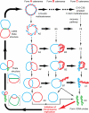
FIGURE 1.
FIGURE 1.
The multicatenane pathway of chromosome segregation (5). Thick horizontal arrows denote pathways of DNA synthesis that convert gapped or nicked DNA circles into covalently closed circles. Thin vertical arrows denote pathways of decatenation of the intertwined daughter chromosomes. Daughter DNA duplexes are in blue and red, whereas parental double-stranded DNA is in green. The monomeric (completely decatenated) and supercoiled daughter circles are also in green, as they become parental DNA in the next round of replication (red arrow). The A, B, and C notations of multicatenated DNA dimers correspond to both circles relaxed (nicked or gapped), one circle covalently closed (and therefore supercoiled), and both circles covalently closed, respectively. Numbers, e.g. A10–A30, are catenation linking numbers, i.e. the levels of intertwining of double helices in the topologically linked circles. The theta structures (replication intermediates) in brackets are diagrams of thetas in which the parental (green) DNA is nicked and therefore relaxed as histone-free DNA. The indicated multicatenanes such as B5, C3, C5, etc., are not depicted explicitly, given their highly “entangled” configurations as free DNA. Among the technical innovations in these 1980–1981 studies (–5) was the discovery that hypertonic treatment of SV40-infected green monkey CV1 cells arrested replicated chromosomes at the stage of decatenation. During arrest, highly intertwined nicked (or gapped) A-type multicatenanes were eventually converted (via B-type intermediates) to the equally highly intertwined C-type multicatenanes (C10–C30), in which both circles were covalently closed and supercoiled (thick dashed arrows at the top). The supercoiled state stemmed from the presence of nucleosomes in the minichromosomes, prior to removal of histones for analyses of DNA topology. Upon release from the decatenation block, the highly intertwined C-type multicatenanes were decatenated to supercoiled monomers (vertical dashed arrows; “recovery pathway”). The actual paths taken during chromosome segregation (in the absence of decatenation arrest) include the entire matrix of transitions shown in the diagram. The relative rates of flow along specific paths depend on the rates of gap-filling DNA synthesis (converting a nicked or gapped circle into a covalently closed circle) versus the rates of decatenation of multiply intertwined chromosomes by topoisomerase II (3, 4).
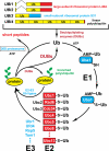
FIGURE 2.
FIGURE 2.
The Ub system of the yeast S. cerevisiae. The fundamental design of this system is conserved among eukaryotes. The yeast Ub genes UBI1–UBI4, two of which contain introns, encode fusions of Ub either to itself or to one of two ribosomal proteins. These fusions are cleaved by DUB enzymes, yielding mature Ub. Thioester bonds between Ub and the active-site Cys residues of Ub-specific enzymes are indicated (∼). The conjugation of Ub to other proteins involves a preliminary ATP-dependent step in which the last residue of Ub (Gly76) is joined, via a thioester bond, to a Cys residue of the E1 (Ub-activating) enzyme, encoded by UBA1. The activated Ub is transferred to a Cys residue in one of several Ub-conjugating (E2) enzymes, encoded by the UBC family genes, and from there to a Lys residue of an ultimate acceptor protein. E2 enzymes function as subunits of E2-E3 Ub ligase holoenzymes. The functions of E3 include the initial recognition of a substrate's degradation signal (degron) and the E2/E3-mediated formation of substrate-linked poly-Ub chains (green ovals). The names of some of the ∼200 E3 enzymes of S. cerevisiae are indicated as well. A targeted ubiquitylated protein is processively degraded to short peptides by the ATP-dependent 26 S proteasome.
FIGURE 3.
FIGURE 3.
The mammalian N-end rule pathway. N-terminal residues are indicated by single-letter abbreviations for amino acids. Yellow ovals denote the rest of a protein substrate. A sign, above hemin in the middle of diagram, is a modified “down-regulation” sign that denotes, specifically, a down-regulation mediated, at least in part, by target's degradation. C* denotes oxidized Cys, either Cys sulfinate or Cys sulfonate, produced in reactions mediated by NO, O2, and their derivatives. Oxidized N-terminal Cys is arginylated by ATE1-encoded isoforms of R-transferase. Type 1 and 2 primary destabilizing N-terminal residues are recognized by the pathway's E3 Ub ligases, called N-recognins. Through their other substrate-binding sites, these E3 enzymes also recognize internal (non-N-terminal) degrons in other substrates of the N-end rule pathway, denoted by the larger oval. As shown in the diagram, hemin (Fe3+-heme) interacts not only with ATE1 (R-transferase) but with UBR family Ub ligases as well, and down-regulates at least some E3 enzymes of this family (89).

References

    1. Varshavsky, A., Sundin, O., and Bohn, M. (1978) Nucleic Acids Res. 5 3469–3478 - PMC - PubMed
    1. Varshavsky, A., Sundin, O., and Bohn, M. (1979) Cell 16 453–466 - PubMed
    1. Sundin, O., and Varshavsky, A. (1980) Cell 21 103–114 - PubMed
    1. Sundin, O., and Varshavsky, A. (1981) Cell 25 659–669 - PubMed
    1. Varshavsky, A., Levinger, L., Sundin, O., Barsoum, J., Özkaynak, E., Swerdlow, P., and Finley, D. (1983) Cold Spring Harb. Symp. Quant. Biol. 47 511–528 - PubMed

Publication types

LinkOut - more resources