. 2008 Nov 15;95(10):4631-42.
doi: 10.1529/biophysj.108.134551. Epub 2008 Aug 15.

Models of toxic beta-sheet channels of protegrin-1 suggest a common subunit organization motif shared with toxic alzheimer beta-amyloid ion channels

Affiliations

Models of toxic beta-sheet channels of protegrin-1 suggest a common subunit organization motif shared with toxic alzheimer beta-amyloid ion channels

Hyunbum Jang et al. Biophys J. .

Abstract

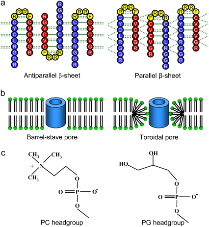
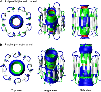
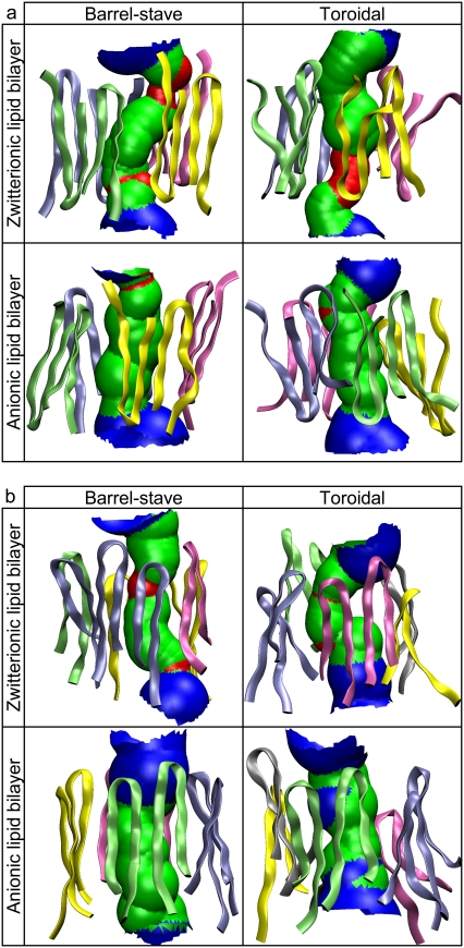
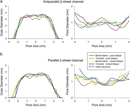
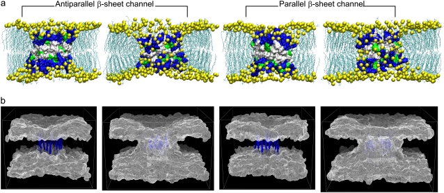
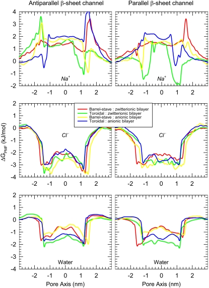
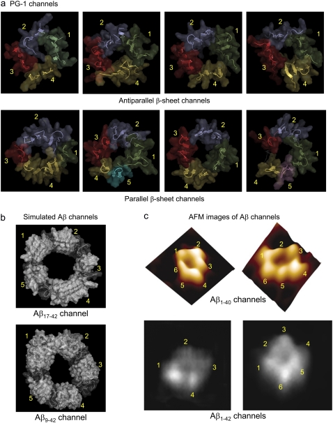
Antimicrobial peptides (AMPs) induce cytotoxicity by altering membrane permeability. The electrical properties of membrane-associated AMPs as well as their cellular effects have been extensively documented; however their three-dimensional structure is poorly understood. Gaining insight into channel structures is important to the understanding of the protegrin-1 (PG-1) and other AMP cytolytic mechanisms, and to antibiotics design. We studied the beta-sheet channels morphology using molecular dynamics simulations. We modeled PG-1 channels as intrinsic barrel-stave and toroidal membrane pores, and simulated them in zwitterionic and anionic lipid bilayers. PG-1 channels consist of eight beta-hairpins in a consecutive NCCN (N and C represent the beta-hairpin's N- and C-termini) packing organization yielding antiparallel and parallel beta-sheet channels. Both channels preserve the toroidal, but not the barrel-stave pores. The two lipid leaflets of the bilayer bend toward each other at the channels' edges, producing a semitoroidal pore with the outward-pointing hydrophobic residues preventing the polar lipid headgroups from moving to the bilayer center. In all simulated lipid environments, PG-1 channels divide into four or five beta-sheet subunits consisting of single or dimeric beta-hairpins. The channel morphology with subunit organization is consistent with the four to five subunits observed by NMR in the POPE/POPG bilayer. Remarkably, a beta-sheet subunit channel motif is in agreement with Alzheimer ion channels modeled using the universal U-shape beta-strand-turn-beta-strand structure, as well as with high resolution atomic force microscopy images of beta-amyloid channels with four to six subunits. Consistent with the toxic beta-amyloid channels that are ion-conducting, the PG-1 channels permeate anions.


Figures

FIGURE 1
FIGURE 1
Topological diagrams (a) for the PG-1 channels in the antiparallel (left) and parallel (right) β-sheet arrangements in an NCCN packing mode. Blue and red represent the N-terminal and C-terminal residues, respectively, and yellow represents the loop residues in the β-hairpin. (b) Cartoon representations of the barrel-stave (left) and toroidal (right) membrane pores. Hollow cylinders represent PG-1 channels. (c) Schematic diagrams of the lipid headgroups for the zwitterionic POPC (left) and anionic POPG (right) lipids.
FIGURE 2
FIGURE 2
Starting points of the channels in a cartoon representation for the (a) antiparallel and (b) parallel β-sheet channels of PG-1. In the peptides, hydrophobic residues are shown in white, one polar (Tyr7) and three Gly (Gly2, Gly3, and Gly17) residues are shown in green, and six positively charged Arg residues (Arg1, Arg4, Arg9, Arg10, Arg11, and Arg18) are shown in blue. In each channel, the initial water pore structure calculated by the HOLE program (43) is embedded. For the pore structures in the surface representation, red denotes pore diameter of d < 0.8 nm, green denotes pore diameter in the range, 0.8 nm ≤ d ≤ 1.2 nm, and blue denotes pore diameter of d > 1.2 nm.
FIGURE 3
FIGURE 3
Channel structures averaged over the simulation in a ribbon representation for the (a) antiparallel and (b) parallel β-sheet channels of PG-1 are shown in the lateral view from the lipid bilayer. Peptides are colored according to the subunit organization in the channels. In each channel, the averaged water pore structure calculated by the HOLE program (43) is embedded. For the pore structures in the surface representation, red denotes pore diameter of d < 0.8 nm, green denotes pore diameter in the range, 0.8 nm ≤ d ≤ 1.2 nm, and blue denotes pore diameter of d > 1.2 nm.
FIGURE 4
FIGURE 4
Averaged outer channel diameter and pore diameters (solid lines) as a function of the distance along the pore center axis for the (a) antiparallel and (b) parallel β-sheet channels of PG-1. Dotted lines represent the initial channel dimension at the starting points.
FIGURE 5
FIGURE 5
Fractions of intermolecular and intramolecular backbone hydrogen bonds (H-bonds), QH-bond, for the (a) antiparallel and (b) parallel β-sheet channels of PG-1. Fractions of intramolecular H-bond in the N-C monomer interface are shown as white symbols under each peptide number, and the fractions of intermolecular H-bond in the N-N and C-C dimer interfaces are shown as gray symbols in between the peptide numbers. The peptides 2-3, 4-5, 6-7, and 8-1 contact with the N-N and dimer interface, whereas the peptides 1-2, 3-4, 5-6, and 7-8 contact with the C-C dimer interface.
FIGURE 6
FIGURE 6
(a) Interaction energies of each monomer in the channels with lipids for the antiparallel (upper) and parallel (lower) β-sheet channels of PG-1. (b) The total interaction energy of the PG-1 channels with lipid can be obtained by adding all monomers interaction energies.
FIGURE 7
FIGURE 7
(a) Snapshots of the channel-bilayer systems at the end of simulations for the PG-1 channels in the anionic lipid bilayer. The first and third snapshots (from left) correspond to the bilayer with the intrinsic barrel-stave membrane pore, and second and fourth snapshots represent the bilayer with the intrinsic toroidal membrane pore for each β-sheet channel. In the surface representations of the channel structures, hydrophobic residues are shown in white, polar and Gly residues are shown in green, and positively charged residues are shown in blue. Phosphates in the lipid headgroups are shown as yellow beads and the lipid tails are shown as threads. Water molecules and ions are removed for clarity. (b) Three-dimensional density maps of the lipid headgroup for the anionic lipid bilayer. The intrinsic barrel-stave membrane pores (the first and third maps from left for the antiparallel and parallel β-sheet channels, respectively) become the semitoroidal membrane pores, whereas the intrinsic toroidal membrane pores (the second and fourth maps from the left for the antiparallel and parallel β-sheet channels, respectively) are well-preserved during the simulations. The density maps represent the angle view of the time averaged bilayer structure. The PG-1 channels in a ribbon representation in blue are embedded in the density maps.
FIGURE 8
FIGURE 8
PMF, ΔGPMF, representing the relative free energy profile for Na+, Cl, and water as a function of the distance along the pore center axis for the antiparallel (left column) and parallel (right column) β-sheet channels of PG-1.
FIGURE 9
FIGURE 9
Side-by-side comparison between the PG-1 and Aβ channels. All channels are viewed from the top leaflet of the lipid bilayer. (a) The simulated PG-1 channels show the antiparallel (Aba-pc, Ato-pc, Aba-pg, and Ato-pg channels from left) and parallel (Pba-pc, Pto-pc, Pba-pg, and Pto-pg channels from left) β-sheet channels. The PG-1 channels are depicted in a cartoon representation with a transparent surface. Each subunit in the channels is colored in a different color. The discontinuous β-sheet network determines the boundary between the subunits in the channels. Antiparallel β-sheet channels of PG-1 contain four subunits. Each subunit is a β-sheet dimer (peptides 1 and 2 (green), peptides 3 and 4 (blue), peptides 5 and 6 (red), and peptides 7 and 8 (yellow)). Parallel β-sheet channels contain four to five subunits. The shapes of the subunits vary, with β-hairpin monomers, and β-sheet dimer or trimer; there are four subunits in the Pba-pc channel (peptide 1 (green), peptides 2–4 (blue), peptide 5 (red), and peptides 6–8 (yellow)), five subunits in the Pto-pc channel (peptides 1–8 (green), peptides 2 and 3 (blue), peptides 4 and 5 (red), peptide 6 (yellow), and peptide 7 (cyan)), four subunits in the Pba-pg channel (peptides 1–8 (green), peptides 2 and 3 (blue), peptides 4 and 5 (red), and peptides 6 and 7 (yellow)), and five subunits in the Pto-pg channel (peptides 1–8 (green), peptides 2 and 3 (blue), peptide 4 (red), peptide 6 (yellow), and peptide 7 (magenta); here peptide 5 is disordered). (b) The simulated channels show the Aβ17-42 and Aβ9-42 channels (24-mer). (Taken from Jang et al. (32). Permission obtained.) (c) AFM images show the Aβ1-40 and Aβ1-42 channels. (Taken from Quist et al. (26) and Lin et al. (27). Permission obtained.)

Similar articles

Cited by

References

    1. Sitaram, N., and R. Nagaraj. 2002. Host-defense antimicrobial peptides: importance of structure for activity. Curr. Pharm. Des. 8:727–742. - PubMed
    1. Hancock, R. E., and A. Rozek. 2002. Role of membranes in the activities of antimicrobial cationic peptides. FEMS Microbiol. Lett. 206:143–149. - PubMed
    1. Kourie, J. I., and A. A. Shorthouse. 2000. Properties of cytotoxic peptide-formed ion channels. Am. J. Physiol. Cell Physiol. 278:C1063–C1087. - PubMed
    1. Zasloff, M. 2002. Antimicrobial peptides of multicellular organisms. Nature. 415:389–395. - PubMed
    1. Sokolov, Y., T. Mirzabekov, D. W. Martin, R. I. Lehrer, and B. L. Kagan. 1999. Membrane channel formation by antimicrobial protegrins. Biochim. Biophys. Acta. 1420:23–29. - PubMed

Publication types

LinkOut - more resources