

Simulations of skin barrier function: free energies of hydrophobic and hydrophilic transmembrane pores in ceramide bilayers
- PMID: 18708461
- PMCID: PMC2576386
- DOI: 10.1529/biophysj.108.138545
Simulations of skin barrier function: free energies of hydrophobic and hydrophilic transmembrane pores in ceramide bilayers
Abstract
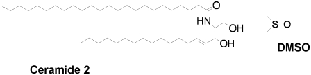
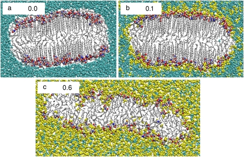
Transmembrane pore formation is central to many biological processes such as ion transport, cell fusion, and viral infection. Furthermore, pore formation in the ceramide bilayers of the stratum corneum may be an important mechanism by which penetration enhancers such as dimethylsulfoxide (DMSO) weaken the barrier function of the skin. We have used the potential of mean constraint force (PMCF) method to calculate the free energy of pore formation in ceramide bilayers in both the innate gel phase and in the DMSO-induced fluidized state. Our simulations show that the fluid phase bilayers form archetypal water-filled hydrophilic pores similar to those observed in phospholipid bilayers. In contrast, the rigid gel-phase bilayers develop hydrophobic pores. At the relatively small pore diameters studied here, the hydrophobic pores are empty rather than filled with bulk water, suggesting that they do not compromise the barrier function of ceramide membranes. A phenomenological analysis suggests that these vapor pores are stable, below a critical radius, because the penalty of creating water-vapor and tail-vapor interfaces is lower than that of directly exposing the strongly hydrophobic tails to water. The PMCF free energy profile of the vapor pore supports this analysis. The simulations indicate that high DMSO concentrations drastically impair the barrier function of the skin by strongly reducing the free energy required for pore opening.
Figures


Similar articles
-
The permeability enhancing mechanism of DMSO in ceramide bilayers simulated by molecular dynamics.Biophys J. 2007 Sep 15;93(6):2056-68. doi: 10.1529/biophysj.107.104703. Epub 2007 May 18. Biophys J. 2007. PMID: 17513383 Free PMC article.
-
The importance of membrane defects-lessons from simulations.Acc Chem Res. 2014 Aug 19;47(8):2244-51. doi: 10.1021/ar4002729. Epub 2014 Jun 3. Acc Chem Res. 2014. PMID: 24892900
-
Ion leakage through transient water pores in protein-free lipid membranes driven by transmembrane ionic charge imbalance.Biophys J. 2007 Mar 15;92(6):1878-90. doi: 10.1529/biophysj.106.094797. Epub 2007 Jan 5. Biophys J. 2007. PMID: 17208976 Free PMC article.
-
How Lipid Membranes Affect Pore Forming Toxin Activity.Acc Chem Res. 2015 Dec 15;48(12):3073-9. doi: 10.1021/acs.accounts.5b00403. Epub 2015 Dec 7. Acc Chem Res. 2015. PMID: 26641659 Review.
-
Structure of the skin barrier and its modulation by vesicular formulations.Prog Lipid Res. 2003 Jan;42(1):1-36. doi: 10.1016/s0163-7827(02)00028-0. Prog Lipid Res. 2003. PMID: 12467638 Review.
Cited by
-
Effect of Ceramide Tail Length on the Structure of Model Stratum Corneum Lipid Bilayers.Biophys J. 2018 Jan 9;114(1):113-125. doi: 10.1016/j.bpj.2017.10.031. Biophys J. 2018. PMID: 29320678 Free PMC article.
-
Improved Topical Drug Delivery: Role of Permeation Enhancers and Advanced Approaches.Pharmaceutics. 2022 Dec 15;14(12):2818. doi: 10.3390/pharmaceutics14122818. Pharmaceutics. 2022. PMID: 36559311 Free PMC article. Review.
-
In Silico Prediction of Stratum Corneum Partition Coefficients via COSMOmic and Molecular Dynamics Simulations.J Phys Chem B. 2023 Mar 30;127(12):2719-2728. doi: 10.1021/acs.jpcb.2c08566. Epub 2023 Mar 17. J Phys Chem B. 2023. PMID: 36930176 Free PMC article.
-
Preparation, Characterization and ex vivo Skin Permeability Evaluation of Type I Collagen-Loaded Liposomes.Int J Nanomedicine. 2023 Apr 6;18:1853-1871. doi: 10.2147/IJN.S404494. eCollection 2023. Int J Nanomedicine. 2023. PMID: 37057190 Free PMC article.
-
Simulation study of the structure and phase behavior of ceramide bilayers and the role of lipid head group chemistry.J Chem Theory Comput. 2013 Nov 12;9(11):5116-5126. doi: 10.1021/ct400431e. J Chem Theory Comput. 2013. PMID: 24501589 Free PMC article.
References
-
- Cohen, F. S., and G. B. Melikyan. 2004. The energetics of membrane fusion from binding, through hemifusion, pore formation, and pore enlargement. J. Membr. Biol. 199:1–14. - PubMed
-
- Cevc, G. 2004. Lipid vesicles and other colloids as drug carriers on the skin. Adv. Drug Deliv. Rev. 56:675–711. - PubMed
-
- Tieleman, D. P. 2006. Computer simulations of transport through membranes: passive diffusion, pores, channels and transporters. Clin. Exp. Pharmacol. Physiol. 33:893–903. - PubMed
-
- Javadov, S., and M. Karmazyn. 2007. Mitochondrial permeability transition pore opening as an endpoint to initiate cell death and as a putative target for cardioprotection. Cell. Physiol. Biochem. 20:1–22. - PubMed
-
- Tolpekina, T. V., W. K. den Otter, and W. J. Briels. 2004. Simulations of stable pores in membranes: system size dependence and line tension. J. Chem. Phys. 121:8014–8020. - PubMed
Publication types
MeSH terms
Substances
LinkOut - more resources
Full Text Sources
Other Literature Sources
