. 2008 Nov 15;95(10):4837-44.
doi: 10.1529/biophysj.108.140061. Epub 2008 Aug 15.

Neither helix in the coiled coil region of the axle of F1-ATPase plays a significant role in torque production

Affiliations

Neither helix in the coiled coil region of the axle of F1-ATPase plays a significant role in torque production

Mohammad Delawar Hossain et al. Biophys J. .

Abstract

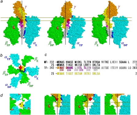
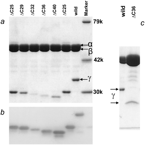
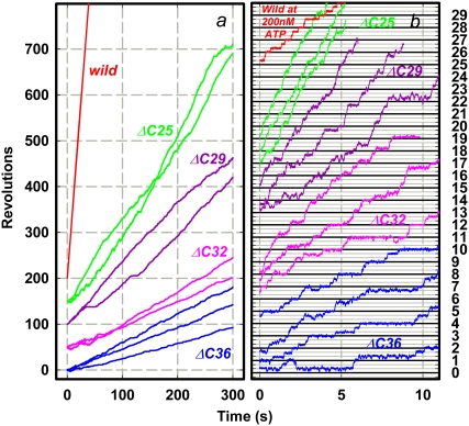
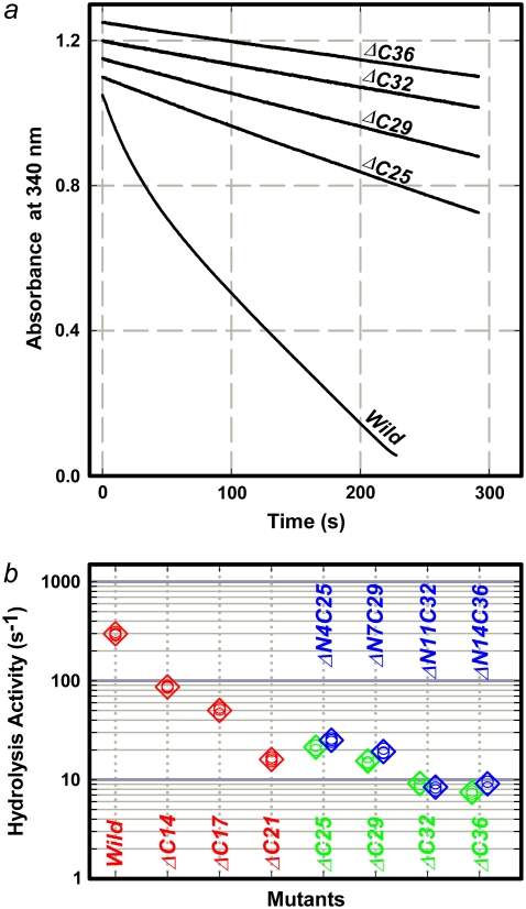
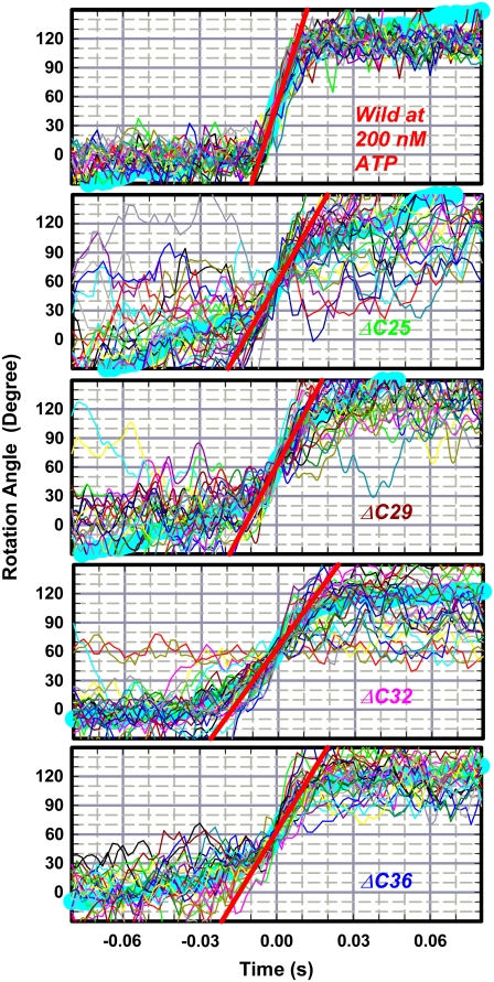
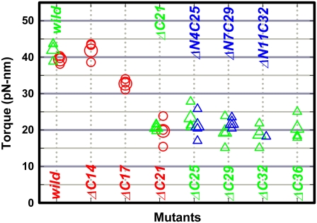
F(1)-ATPase is an ATP-driven rotary molecular motor in which the central gamma-subunit rotates inside the cylinder made of alpha(3)beta(3) subunits. The amino and carboxy termini of the gamma-subunit form the axle, an alpha-helical coiled coil that deeply penetrates the stator cylinder. We previously truncated the axle step by step, starting with the longer carboxy terminus and then cutting both termini at the same levels, resulting in a slower yet considerably powerful rotation. Here we examine the role of each helix by truncating only the carboxy terminus by 25-40 amino-acid residues. Longer truncation impaired the stability of the motor complex severely: 40 deletions failed to yield rotating the complex. Up to 36 deletions, however, the mutants produced an apparent torque at nearly half of the wild-type torque, independent of truncation length. Time-averaged rotary speeds were low because of load-dependent stumbling at 120 degrees intervals, even with saturating ATP. Comparison with our previous work indicates that half the normal torque is produced at the orifice of the stator. The very tip of the carboxy terminus adds the other half, whereas neither helix in the middle of the axle contributes much to torque generation and the rapid progress of catalysis. None of the residues of the entire axle played a specific decisive role in rotation.


Figures

FIGURE 1
FIGURE 1
Atomic structure of MF1 (22). Truncations of γ-subunit are shown, with color scheme in c; the N-terminal α-helix is shown in yellow. Those atoms of α-subunits and β-subunits that are within 0.5 nm from an atom of γ (excluding hydrogens) are colored blue and dark green, respectively. Nucleotides are shown in CPK colors. Black lines in side views and black dots in bottom views represent putative rotation axis (21). (a) Side views show central γ and an opposing α-β pair. Membrane-embedded F0 portion of ATP synthase would be above the γ-subunit. (b) Bottom view of section between pair of gold lines in a. (c) Amino-acid sequences at the C-terminus and N-terminus of γ in MF1 (36) and TF1 (30), except that numbering for TF1 in our study here starts from Met-1, which is absent in the expressed wild-type protein. In Fig. 1 B of Furuike et al. (27), Ala-2 was incorrectly shown as Ala-1. (d) Central portions of bottom and side views for γ-ΔC36.
FIGURE 2
FIGURE 2
Confirmation of γ-truncations by polyacrylamide gel electrophoresis. (a) Ten percent gel contains 0.1% SDS, stained with Coomassie brilliant Blue R-250. (b) Western blot of a stained with anti-γ-antibody. (c) Overloaded gel shows absence of intact γ in mutant. Amount of γ in purified samples was variable, depending on preparations; γ-ΔC40 in another preparation showed a barely detectable γ band. Side bands in b are presumably dissociated and degraded γ.
FIGURE 3
FIGURE 3
Time courses of rotation of 0.29-μm bead duplex attached to γ-subunit. All rotations were counterclockwise when viewed from above in Fig. 1 a. Horizontal lines in b are separated by 120°. ATP concentration was 2 mM, except for the wild-type in b, which stepped because of infrequent binding of ATP at 200 nM (rate constant for ATP binding (13), ∼2 × 10⁷ M−1s−1). Dwelling angles of mutants are plotted ∼80° ahead of ATP-waiting dwells of the wild-type, because long pauses such as those in γ-ΔC29 in a also occurred at same angles and because long pauses in the wild-type, because of MgADP inhibition (11), occur at ∼80°. This assignment, however, is tentative because we could not determine ATP-waiting angles of mutants. Bottom curve in b shows an exceptional case of reversal over two 120°-steps in a sluggish phase, after which more regular rotation resumed beyond the right-hand axis. Temperature, 23°C.
FIGURE 4
FIGURE 4
ATP hydrolysis activity. (a) Time courses of hydrolysis monitored as decrease in NADH absorbance at 340 nm. A decrease of one absorbance unit corresponds to hydrolysis of 1.6 × 10⁻⁴ M of ATP. Reaction was initiated by adding F1 at a final concentration of 10 nM (mutants) or 5 nM (wild-type) to coupled-assay medium containing 2 mM ATP. (b) Summary of hydrolysis rates at 2 mM ATP. Green, this work; red, previous work (26), in which mutants were prepared with a regular procedure including heat shock, and bound nucleotides were removed; blue, simultaneous truncation of both N-terminus and C terminus of γ-subunit (27), where treatment with size-exclusion column was performed at room temperature, which helped to stabilize mutants. Small circles indicate individual data, and large diamonds indicate their average.
FIGURE 5
FIGURE 5
Stepping records for torque estimation. Thin colored curves show 30 consecutive steps; thick cyan curve constitutes their average. Individual step records were shifted vertically by a multiple of 120° to obtain overlap. Time zero for each step record was assigned by eye to the data point closest to 60°. Straight red lines indicate linear fit to the cyan curve between 30° and 90°.
FIGURE 6
FIGURE 6
Torque of gamma deletion mutants. Green, this study; red (26) and blue (27), previous results. Circles, torque estimated with a duplex of 0.49-μm beads; triangles, 0.29-μm bead duplex. Small symbols represent individual estimations, and large symbols represent their average.

Similar articles

Cited by

References

    1. Kagawa, Y., and E. Racker. 1966. Partial resolution of the enzymes catalyzing oxidative phosphorylation. IX. Reconstruction of oligomycin-sensitive adenosine triphosphatase. J. Biol. Chem. 241:2467–2474. - PubMed
    1. Catteral, W. A., and P. L. Pedersen. 1971. Adenosine triphosphatase from rat liver mitochondria. I. Purification, homogeneity, and physical properties. J. Biol. Chem. 246:4987–4994. - PubMed
    1. Cox, G. B., J. A. Downie, L. Langman, A. E. Senior, G. Ash, D. R. H. Fayle, and F. Gibson. 1981. Assembly of the adenosine triphosphatase complex in Escherichia coli: assembly of F0 is dependent on the formation of specific F1 subunits. J. Bacteriol. 148:30–42. - PMC - PubMed
    1. Yoshida, M., E. Muneyuki, and T. Hisabori. 2001. ATP synthase—a marvelous rotary engine of the cell. Nat. Rev. Mol. Cell Biol. 2:669–677. - PubMed
    1. Boyer, P. D., and W. E. Kohlbrenner. 1981. The present status of the binding-change mechanism and its relation to ATP formation by chloroplasts. In Energy Coupling in Photosynthesis. B. R. Selman and S. Selman-Reimer, editors. Elsevier, Amsterdam, The Netherlands. 231–240.

Publication types

MeSH terms

Substances

LinkOut - more resources