. 2008 Aug;20(8):2252-64.
doi: 10.1105/tpc.108.058685. Epub 2008 Aug 15.

RD19, an Arabidopsis cysteine protease required for RRS1-R-mediated resistance, is relocalized to the nucleus by the Ralstonia solanacearum PopP2 effector

Affiliations

RD19, an Arabidopsis cysteine protease required for RRS1-R-mediated resistance, is relocalized to the nucleus by the Ralstonia solanacearum PopP2 effector

Maud Bernoux et al. Plant Cell. 2008 Aug.

Erratum in

  • CORRECTION.
    [No authors listed] [No authors listed] Plant Cell. 2020 Sep;32(9):3036-3037. doi: 10.1105/tpc.20.00482. Epub 2020 Jul 2. Plant Cell. 2020. PMID: 32616660 Free PMC article. No abstract available.

Abstract

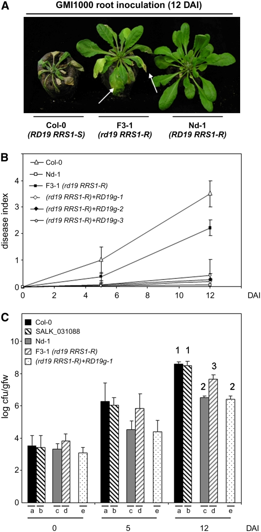
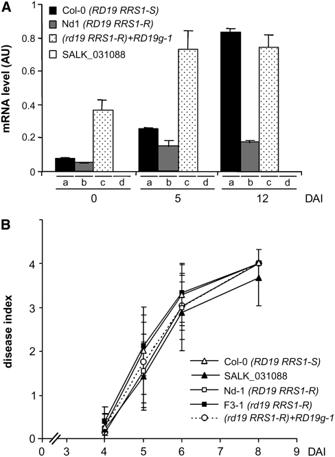
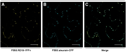
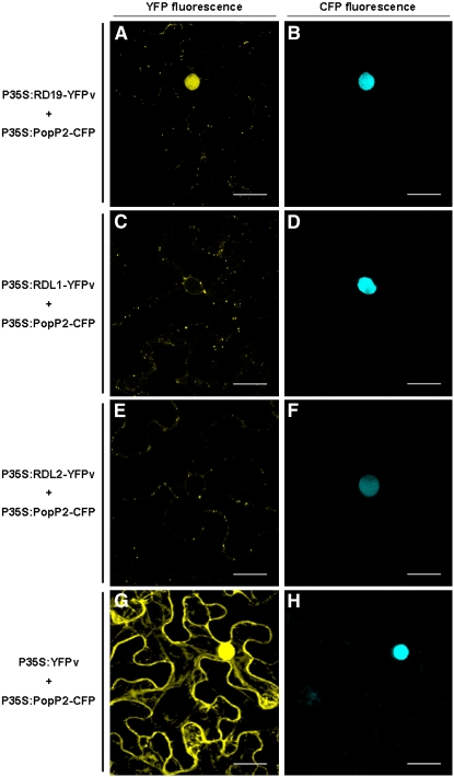
Bacterial wilt, a disease impacting cultivated crops worldwide, is caused by the pathogenic bacterium Ralstonia solanacearum. PopP2 (for Pseudomonas outer protein P2) is an R. solanacearum type III effector that belongs to the YopJ/AvrRxv protein family and interacts with the Arabidopsis thaliana RESISTANT TO RALSTONIA SOLANACEARUM 1-R (RRS1-R) resistance protein. RRS1-R contains the Toll/Interleukin1 receptor-nucleotide binding site-Leu-rich repeat domains found in several cytoplasmic R proteins and a C-terminal WRKY DNA binding domain. In this study, we identified the Arabidopsis Cys protease RESPONSIVE TO DEHYDRATION19 (RD19) as being a PopP2-interacting protein whose expression is induced during infection by R. solanacearum. An Arabidopsis rd19 mutant in an RRS1-R genetic background is compromised in resistance to the bacterium, indicating that RD19 is required for RRS1-R-mediated resistance. RD19 normally localizes in mobile vacuole-associated compartments and, upon coexpression with PopP2, is specifically relocalized to the plant nucleus, where the two proteins physically interact. No direct physical interaction between RRS1-R and RD19 in the presence of PopP2 was detected in the nucleus as determined by Förster resonance energy transfer. We propose that RD19 associates with PopP2 to form a nuclear complex that is required for activation of the RRS1-R-mediated resistance response.


Figures

Figure 1.
Figure 1.
RD19 Is Required for RRS1-R–Mediated Disease Resistance Signaling. (A) Phenotypic responses of susceptible Col-0 (RD19 RRS1-S) and resistant Nd-1 (RD19 RRS1-R) wild-type Arabidopsis plants and of the rd19 RRS1-R (F3-1) line to the GMI1000 strain of R. solanacearum 12 DAI. Arrows point to wilted leaves of the mutant line. (B) Disease symptom development curves. Each plant was scored at 0, 5, and 12 DAI of roots using a scale between 0 and 4: 0 = no wilting, 1 = 25%, 2 = 50%, 3 = 75%, and 4 =100% of leaves were wilted. Means and sd were calculated from scores of a total of 60 plants per genotype (from three independent experiments); triangle, Col-0 (RD19 RRS1-S); open square, Nd-1(RD19 RRS1-R); closed square, rd19 mutant (F3-1 line [rd19 RRS1-R]); open diamonds, closed diamond, and open circle, three independent complemented rd19 lines (RD19g1 to -3 in an rd19 RRS1-R background). (C) Bacterial growth inside the plant was estimated as described before (Deslandes et al., 1998). Means of colony-forming units per gram of fresh weight (cfu/gfw) and sd were calculated from triplicates of three plants for each genotype (from three independent experiments). (a) Col-0 (RD19 RRS1-S), (b) SALK_031088 line (rd19 RRS1-S), (c) Nd-1(RD19 RRS1-R), (d) rd19 RRS1-R F3-1 line, (e) complemented rd19 line (RD19g1 in an rd19 RRS1-R background). Analysis of variance and multiple between-group comparisons (Bonferroni test) were used to analyze differences between ecotypes. Groups 1 (a and b), 2 (d), and 3 (c and e) are statistically different (P value of 0.05).
Figure 2.
Figure 2.
Enhanced Expression of RD19 Does Not Affect the Plant Response to the ΔPopP2 Strain. (A) Expression analysis of the RD19 gene in three different genotypes after inoculation with the GMI1000 strain. RD19 transcript levels were determined by Q-RT-PCR from cDNAs generated from the aerial parts of three plants per genotype at 0, 5, and 12 DAI. The expression values of RD19 were normalized using the expression level of two housekeeping genes considered as internal standards. Mean expression and sd values were calculated from the results of two independent experiments (triplicate samples of three plants were taken at each time point). (a) Col-0 (RD19 RRS1-S), (b) Nd-1(RD19 RRS1-R), (c) RD19g-1 line (rd19 RRS1-R mutant complemented with RD19 genomic clone), (d) rd19 RRS1-S mutant (SALK_031088). AU, arbitrary units. (B) Disease symptom developments were scored after root inoculation with the ΔPopP2 strain. Each plant was scored at 4, 5, 6, and 8 DAI of roots using a scale between 0 and 4: 0 = no wilting, 1 = 25%, 2 = 50%, 3 = 75%, and 4 =100% of leaves were wilted. Means and sd were calculated from scores of 30 plants per accession and mutant line. This experiment was repeated three times, and reproducible results were obtained; open triangle, Col-0 (RD19 RRS1-S); open square, Nd-1 (RD19 RRS1-R); closed triangle, SALK_031088 line (rd19 RRS1-S); closed square, F3-1 line (rd19 RRS1-R); open circle, RD19g-1 line (rd19 RRS1-R mutant complemented with RD19 genomic clone).
Figure 3.
Figure 3.
Expression Analysis of PR Genes PR-3, PR-4, and PDF1.2. Transcript levels of PR-3, PR-4, and PDF1.2 genes were determined by Q-RT-PCR from cDNAs generated from aerial parts of three plants per genotype at 0, 5, and 12 DAI. The expression values of the genes were normalized using the expression level of two housekeeping genes considered as internal standards. Mean expression and sd values were calculated from the results of two independent experiments (triplicate samples of three plants were taken at each time point). (a) Col-0 (RD19 RRS1-S), (b) Nd-1(RD19 RRS1-R), (c) rd19 mutant (F3-1 line, rd19 RRS1-R), (d) RD19g-1 line (rd19 RRS1-R mutant complemented with RD19).
Figure 4.
Figure 4.
Subcellular Localization of RD19-YFPv in N. benthamiana. Confocal images of N. benthamiana epidermal cells 48 h after coexpression of P35S:RD19-YFPv and P35S:aleurain-CFP via A. tumefaciens infiltration. RD19-YFPv and aleurain-CFP colocalize in mobile vesicles ([A] and [B], respectively), moving along the endoplasmic reticulum. Colocalization of RD19-YFPv and aleurain-CFP is shown in the merged image in (C). Bars = 20 μm.
Figure 5.
Figure 5.
RD19 Relocalizes to the Plant Nucleus in the Presence of PopP2. Coexpression of RD19-YFPv with PopP2-CFP in N. benthamiana epidermal cells resulted in the relocalization of RD19 into the nucleus ([A] and [B]). YFP fluorescence was not detected in the nucleus when PopP2-CFP was coexpressed with RDL1-YFPv or RDL2-YFPv ([C] and [D] and [E] and [F], respectively). YFP fluorescence was detected in the whole cell when YFPv alone was expressed in the presence of PopP2-CFP ([G] and [H]). Bars = 20 μm.

Similar articles

Cited by

References

    1. Axtell, M.J., and Staskawicz, B.J. (2003). Initiation of RPS2-specified disease resistance in Arabidopsis is coupled to the AvrRpt2-directed elimination of RIN4. Cell 112 369–377. - PubMed
    1. Barrett, A.J., and Rawlings, N.D. (2001). Evolutionary lines of Cysteine peptidases. Biol. Chem. 382 727–733. - PubMed
    1. Belkhadir, Y., Nimchuk, Z., Hubert, D.A., Mackey, D., and Dangl, J.L. (2004). Arabidopsis RIN4 negatively regulates disease resistance mediated by RPS2 and RPM1 downstream or independent of the NDR1 signal modulator and is not required for the virulence functions of bacterial type III effectors AvrRpt2 or AvrRpm1. Plant Cell 16 2822–2835. - PMC - PubMed
    1. Biener, E., Charlier, M., Ramanujan, V.K., Daniel, N., Eisenberg, A., Bjorbaek, C., Herman, B., Gertler, A., and Djiane, J. (2005). Quantitative FRET imaging of leptin receptor oligomerization kinetics in single cells. Biol. Cell 97 905–919. - PubMed
    1. Burch-Smith, T.M., Schiff, M., Caplan, J.L., Tsao, J., Czymmek, K., and Dinesh-Kumar, S.P. (2007). A novel role for the TIR domain in association with pathogen-derived elicitors. PLoS Biol. 5 e68. - PMC - PubMed

Publication types

MeSH terms