. 2008 Oct;190(20):6795-804.
doi: 10.1128/JB.00401-08. Epub 2008 Aug 15.

O2- and NO-sensing mechanism through the DevSR two-component system in Mycobacterium smegmatis

Affiliations

O2- and NO-sensing mechanism through the DevSR two-component system in Mycobacterium smegmatis

Jin-Mok Lee et al. J Bacteriol. 2008 Oct.

Abstract

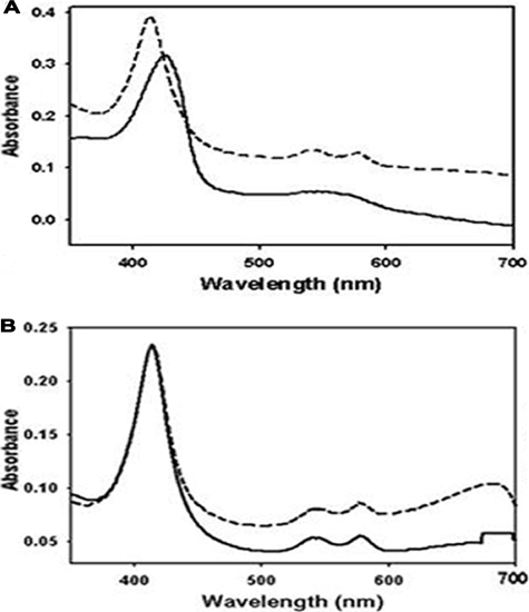
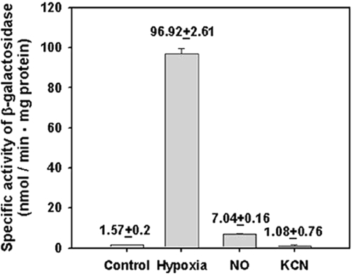
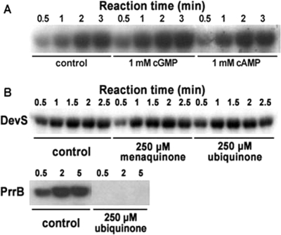
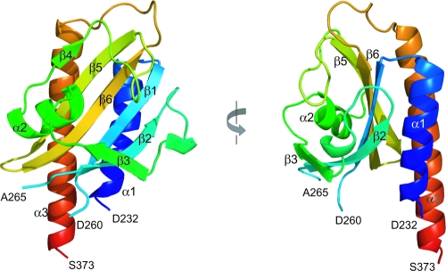
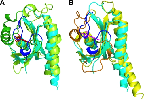
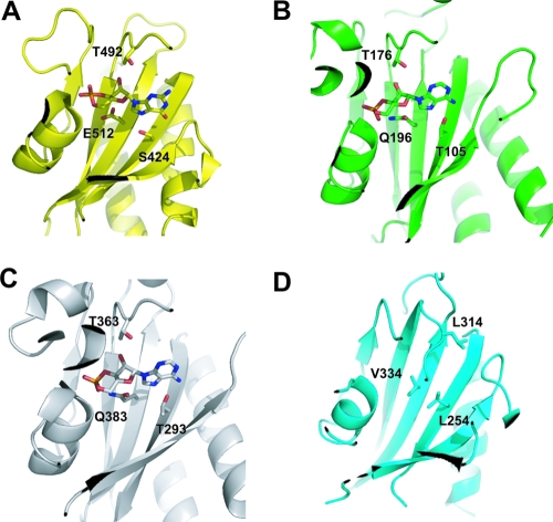
The DevS histidine kinase of Mycobacterium smegmatis contains tandem GAF domains (GAF-A and GAF-B) in its N-terminal sensory domain. The heme iron of DevS is in the ferrous state when purified and is resistant to autooxidation from a ferrous to a ferric state in the presence of O(2). The redox property of the heme and the results of sequence comparison analysis indicate that DevS of M. smegmatis is more closely related to DosT of Mycobacterium tuberculosis than DevS of M. tuberculosis. The binding of O(2) to the deoxyferrous heme led to a decrease in the autokinase activity of DevS, whereas NO binding did not. The regulation of DevS autokinase activity in response to O(2) and NO was not observed in the DevS derivatives lacking its heme, indicating that the ligand-binding state of the heme plays an important role in the regulation of DevS kinase activity. The redox state of the quinone/quinol pool of the respiratory electron transport chain appears not to be implicated in the regulation of DevS activity. Neither cyclic GMP (cGMP) nor cAMP affected DevS autokinase activity, excluding the possibility that the cyclic nucleotides serve as the effector molecules to modulate DevS kinase activity. The three-dimensional structure of the putative GAF-B domain revealed that it has a GAF folding structure without cyclic nucleotide binding capacity.


Figures

FIG. 1.
FIG. 1.
Absorption spectra of purified DevS. The spectra were recorded using 300 μg of DevS in 20 mM Tris-HCl buffer at RT. The DevS protein was purified in the absence of any reducing reagent in the buffer used during the purification procedures. (A) DevS was reduced by the addition of DTT to a final concentration of 10 mM and sparged with N2 gas for 10 min to yield a deoxyferrous derivative of DevS (solid line). The deoxyferrous form of DevS was exposed to air to generate the oxyferrous form of DevS (dashed line). (B) The spectra of the purified DevS before (solid line) and after (dashed line) 10 mM KCN treatment.
FIG. 2.
FIG. 2.
Kinase activities of the DTT-reduced ferrous DevS derivatives. Purified DevS was treated with 10 mM DTT and sparged with N2 gas for 10 min to generate the deoxyferrous form of DevS. Oxy- and NO-ferrous DevS derivatives were prepared as described in Materials and Methods. The autophosphorylation reactions were performed by using 80 pmol each of DevS, C-DevS, and H150A DevS and 200 mM (1,000 ci/mol) ATP in the anaerobic box at RT. At the time points indicated, samples (10 μl) were removed and added to 3 μl of loading buffer to stop the reaction. (A) The phosphorylation of the protein was assayed by SDS-polyacrylamide gel electrophoresis and subsequent autoradiography. (B) Quantitation of the band intensity of phosphorylated DevS was performed with a densitometer program, ImageJ (version 1.37), and the relative values are plotted as a function of the reaction time. Symbols: filled circle, oxyferrous DevS; open circle, deoxyferrous DevS; filled inverted triangle, NO-ferrous DevS.
FIG. 3.
FIG. 3.
Expression of hspX in M. smegmatis grown under various stress conditions: Control, the M. smegmatis strain carrying the hspX::lacZ transcriptional fusion plasmid pNChspX was grown aerobically in 7H9-glucose medium to an OD600 of 0.5; Hypoxia, the strain was grown in a 250-ml flask filled with 150 ml of 7H9-glucose medium and sealed with a rubber septum on a gyratory shaker (200 rpm) for 15 h following inoculation of the medium with aerobically grown preculture, to an OD600 of 0.05; NO and KCN, the strains were initially grown aerobically in 7H9-glucose medium to an OD600 of 0.3. The cultures were treated with either 500 μM sodium nitroprusside or 500 μM KCN and further grown aerobically for an additional 2 h. Cell-free crude extracts were used to determine β-galactosidase activity. The β-galactosidase activity is expressed as nanomoles per minute per milligram of protein. All values provided are the average of the results of two independent determinations. Error bars indicate standard deviations.
FIG. 4.
FIG. 4.
Effects of cyclic nucleotides, menaquinone, and ubiquinone on DevS autokinase activity. (A) The autophosphorylation reactions were performed by using purified DevS (80 pmol) and 200 μM (1,000 ci/mol) ATP at 30°C. The reaction mixtures with either 1 mM cGMP or 1 mM cAMP were incubated on ice for 1 h before the addition of ATP. As a control, the reaction mixture without a cyclic nucleotide was included in the experiment. At the time points indicated, samples (10 μl) were removed and added to 3 μl of loading buffer to stop the reaction. The phosphorylation of the protein was assayed by SDS-polyacrylamide gel electrophoresis and subsequent autoradiography. (B) The autophosphorylation reactions were performed by using 80 pmol each of DevS or PrrB and 100 μM (1,000 Ci/mol) ATP at 30°C. The reaction mixtures with either 250 mM menaquinone or 250 μM ubiquinone were incubated at 30°C for 25 min before the addition of ATP. As a control, the reaction mixture without a quinone compound was included in the experiment.
FIG. 5.
FIG. 5.
Ribbon diagrams of the GAF-B domain of DevS. The domain consists of a six-stranded antiparallel β-sheet and three α-helices. Residues P261 to S264 are missing in the structure. The left diagram is rotated 90° about the vertical axis.
FIG. 6.
FIG. 6.
Structural comparison of the DevS GAF-B domain with GAF domains containing cyclic nucleotides. The structure of the GAF-B domain of DevS (cyan) is superimposed on the structures of the GAF-A domain (green) of adenylyl cyclase CyaB2 (A) and the GAF-B domain (yellow) of PDE2A (B). For the binding of cAMP (red) or cGMP (magenta), the α3-helix and the loop between the β2- and β3-strands of the GAF domains of PDE2A (orange) and CyaB2 (dark green) form the binding pocket. In GAF-B of DevS, two loops corresponding to the orange and dark green regions are presented in blue.
FIG. 7.
FIG. 7.
The multiple sequence alignment of DevS of M. smegmatis with its homologs in M. tuberculosis. Amino acid sequences of DevS from M. smegmatis (Msm_DevS) and DosT (Mtb_DosT) and DevS (Mtb_DevS) from M. tuberculosis were aligned. Numbering was done using DevS of M. smegmatis. The arrows and coils above the aligned sequences indicate the elements of the secondary structure of the DevS GAF-B domain. Conserved residues are shown by the gray background. The putative transmembrane motifs, which were predicted with M. tuberculosis DevS, are denoted by thick lines below the alignment. The amino acid residues corresponding to those hydrogen bonded to a cyclic nucleotide in PDE2A and CyaB2 are indicated by the inverted triangles. Multiple alignment was performed using ClustalW2 (http://www.ebi.ac.uk/Tools/clustalw2/) and visualized using ESPript (http://espript.ibcp.fr/ESPript/ESPript/).
FIG. 8.
FIG. 8.
Hydrophilic residues involved in the binding of cyclic nucleotides in the GAF domains. The residues presented on the β-sheets and the loop linked to the β5 strand are hydrogen bonded directly to the cyclic nucleotides in the GAF-B domain of PDE2A (S424, T492, and E512) (A) and the GAF-A domain (T105, T176, and Q196) (B) and GAF-B domain (T293, T363, and Q383) (C) of adenylyl cyclase CyaB2. These hydrophilic residues are replaced by hydrophobic ones in the GAF-B domain of DevS (L254, V334, and L314) (D).

References

    1. Aly, S., K. Wagner, C. Keller, S. Malm, A. Malzan, S. Brandau, F. C. Bange, and S. Ehlers. 2006. Oxygen status of lung granulomas in Mycobacterium tuberculosis-infected mice. J. Pathol. 210298-305. - PubMed
    1. Aravind, L., and C. P. Ponting. 1997. The GAF domain: an evolutionary link between diverse phototransducing proteins. Trends Biochem. Sci. 22458-459. - PubMed
    1. Berman, H., K. Henrick, and H. Nakamura. 2003. Announcing the worldwide Protein Data Bank. Nat. Struct. Biol. 10980. - PubMed
    1. Charbonneau, H., R. K. Prusti, H. LeTrong, W. K. Sonnenburg, P. J. Mullaney, K. A. Walsh, and J. A. Beavo. 1990. Identification of a noncatalytic cGMP-binding domain conserved in both the cGMP-stimulated and photoreceptor cyclic nucleotide phosphodiesterases. Proc. Natl. Acad. Sci. USA 87288-292. - PMC - PubMed
    1. Dasgupta, N., V. Kapur, K. K. Singh, T. K. Das, S. Sachdeva, K. Jyothisri, and J. S. Tyagi. 2000. Characterization of a two-component system, devR-devS, of Mycobacterium tuberculosis. Tuberc. Lung Dis. 80141-159. - PubMed

Publication types

MeSH terms

LinkOut - more resources