. 2008 Sep 2;105(35):12831-6.
doi: 10.1073/pnas.0805898105. Epub 2008 Aug 15.

Replication termination mechanism as revealed by Tus-mediated polar arrest of a sliding helicase

Affiliations

Replication termination mechanism as revealed by Tus-mediated polar arrest of a sliding helicase

Deepak Bastia et al. Proc Natl Acad Sci U S A. .

Abstract

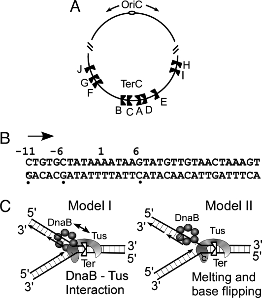
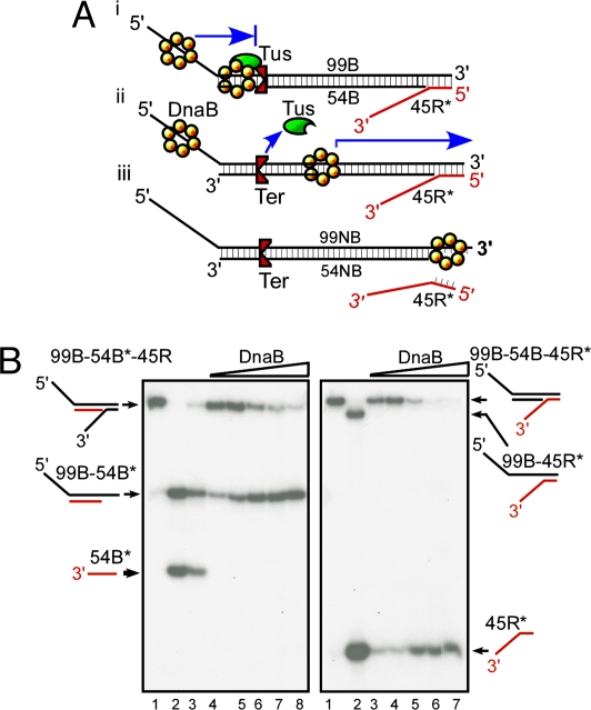
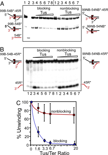
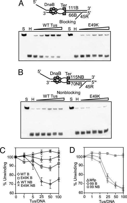
The replication terminator protein Tus of Escherichia coli promotes polar fork arrest at sequence-specific replication termini (Ter) by antagonizing DNA unwinding by the replicative helicase DnaB. Here, we report that Tus is also a polar antitranslocase. We have used this activity as a tool to uncouple helicase arrest at a Tus-Ter complex from DNA unwinding and have shown that helicase arrest occurred without the generation of a DNA fork or a bubble of unpaired bases at the Tus-Ter complex. A mutant form of Tus, which reduces DnaB-Tus interaction but not the binding affinity of Tus for Ter DNA, was also defective in arresting a sliding DnaB. A model of polar fork arrest that proposes melting of the Tus-Ter complex and flipping of a conserved C residue of Ter at the blocking but not the nonblocking face has been reported. The model suggests that enhanced stability of Tus-Ter interaction caused by DNA melting and capture of a flipped base by Tus generates polarity strictly by enhanced protein-DNA interaction. In contrast, the observations presented here show that polarity of helicase and fork arrest in vitro is generated by a mechanism that not only involves interaction between the terminator protein and the arrested enzyme but also of Tus with Ter DNA, without any melting and base flipping in the termination complex.


Conflict of interest statement

The authors declare no conflict of interest.

Figures

Fig. 1.
Fig. 1.
The replication termini of E. coli. (A) Diagram showing the relative locations and orientations of the known Ter sites of E. coli with respect to oriC. (B) The consensus nucleotide sequence of Ter, the C6, and its complementary G are shown. The locations for polar arrest of transcripts catalyzed by E. coli RNA polymerase are shown at −6 and −11. The arrow shows the direction of transcription (and replication). (C) Two models of polar fork arrest. The double-headed arrow in model I indicates Tus–DnaB interaction.
Fig. 2.
Fig. 2.
Experimental strategy for determining DnaB sliding on dsDNA and its arrest at a Tus–Ter complex. (A) Diagram showing the triplex substrates used for measurements of helicase sliding in the blocking orientation that arrests a sliding DnaB and is measured by the unwinding of the 45R reporter oligo (i) and the substrate with the nonblocking orientation of Ter (ii and iii). (B) Phosphorimagergrams showing the products generated by sliding of DnaB on 99B-54B*-45R triplex (Left) and on the 99B-54B-45R* (Right) (* indicates the location of the labeled 5′ end) triplexes in the absence of Tus. The labeled strands are shown in red. Three to four femtomoles of substrate DNA and 0–300 ng of DnaB were used in each reaction. (Left) Lanes 1–3, marker DNA; lanes 4–8, 0, 50, 100, 300, and 400 ng of DnaB, respectively. (Right) Lanes 1 and 2, marker DNA; lanes 3 and 4, DNA without DnaB; and lanes 5–7, 50, 100, and 300 ng of DnaB, respectively.
Fig. 3.
Fig. 3.
Phosphorimagergrams showing the polar arrest of a sliding DnaB without base pair melting by a Tus–Ter complex in the blocking but not in the non-blocking orientation. (A) Representative phosphorimagergrams of products generated by sliding DnaB at a fixed concentration (300 ng) on the 99B-54B*-45R triplex (4 fmole), in the presence of an increasing range of concentration of Tus (molar ratio 0–20) and the same on the 99NB-54NB*-45R triplex. The products are identified by the diagrams on the margins. Blocking Tus; lanes 1–3, marker DNA; lanes 4–8, substrate plus 300 ng DnaB plus Tus at molar ratios of Tus/DNA of 0, 1.6, 3.3, 6.7 and 20 molar. Non blocking Tus; lane 1, marker DNA; lanes 2 and 3, DNA plus 100 and 300 ng of DnaB, respectively; lanes 4–7, DNA plus DnaB (300 ng) plus Tus at molar ratios of, 1.6, 3.3, 6.7, and 20, respectively. B, lanes, 1 and 2, marker DNA; lanes 3–8, DNA substrate plus DnaB (300 ng), plus Tus at molar ratios of 0, 1.6, 3.3, 6.7, 6.7 and 20 respectively.
Fig. 4.
Fig. 4.
Interstrand cross-linking of residues in front of Ter did not abolish arrest of helicase translocation. (A) (Top) Schematic representation of the cross-linked triplex with Ter in the blocking orientation showing the locations of the various oligos and the cross-links (red x). (Middle) Sequence of the triplex with Ter in the blocking orientation about the region of the cross-links; the phenyl selenide substituted oligo is shown in blue except for the GC6 pair that is shown in red with an asterisk; a part of the 30XL sequence is shown in green. (Bottom) Sequence of the control triplex with Ter in the nonblocking orientation; the 26-mer Φ-SeNB oligo sequence is shown in blue except for GC6 that is shown in red with an asterisk; the oligo 20-XL is shown in green; the red X shows the location of the cross-link. (B) The reaction pathway for interstrand T-to-A cross-linking caused by oxidation of an oligonucleotide containing two phenyl-selenide-derivatized T residues. C, autoradiogram of a preparative gel showing the separation of residual noncross-linked 99*-mer from the cross-linked 99*-mer with 24Φ-SeB (arrow). (D) Pooled data from four independent sets of experiments with standard error bars showing the protection of the substrate from melting at the reporter strand by Tus in the blocking and nonblocking orientations of Ter.
Fig. 5.
Fig. 5.
Effects of a mutation in Tus and another in Ter on the arrest of helicase sliding. (A) Representative phosphorimagergrams of gels showing arrest of sliding DnaB by the WT and the E49K mutant form of Tus in the blocking orientation of Ter. S, substrate, H substrate plus DnaB. The wedge indicates addition of an increasing range of molar ratios of Tus to DNA of 1 to 100. (B) The same as A except that the substrate contained the Ter site in the nonblocking orientation. (C) Pooled data from three independent sets of experiments shown with standard error bars. (D) Effect of the transversion GC6 to AT6 in Ter on arrest of DnaB sliding. The triplex was constructed by annealing Mflp1-Mflp2–45R* oligos (Table S1). The data are pooled from eight independent sets of experiments and plotted with standard error bars.

References

    1. Bastia D, Mohanty BK. In: DNA Replication in Eukaryotic Cells. DePamphilis M, editor. Plainview, NY: Cold Spring Harbor Lab Press; 1996. pp. 177–215.
    1. Bastia D, Mohanty B. K. In: DNA Replication and Human Disease. DePamphilis M, editor. Plainview, NY: Cold Spring Harbor Lab Press; 2006. pp. 155–174.
    1. Rothstein R, Michel B, Gangloff S. Replication fork pausing and recombination or “gimme a break”. Genes Dev. 2000;14:1–10. - PubMed
    1. Brewer BJ, Fangman WL. A replication fork barrier at the 3′ end of yeast ribosomal RNA genes. Cell. 1988;55:637–643. - PubMed
    1. Dalgaard JZ, Klar AJ. Orientation of DNA replication establishes mating-type switching pattern in S. pombe. Nature. 1999;400:181–184. - PubMed

Publication types

LinkOut - more resources