. 2008 Aug 15;22(16):2215-27.
doi: 10.1101/gad.1675708.

Identification of cis-acting sites for condensin loading onto budding yeast chromosomes

Affiliations

Identification of cis-acting sites for condensin loading onto budding yeast chromosomes

Claudio D'Ambrosio et al. Genes Dev. .

Abstract

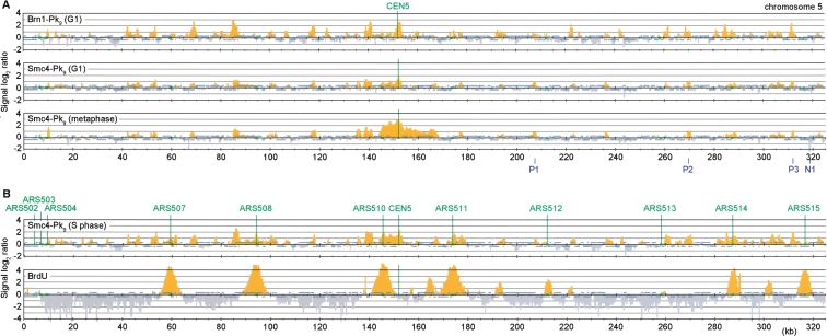
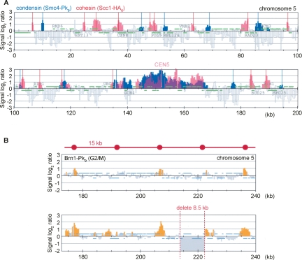
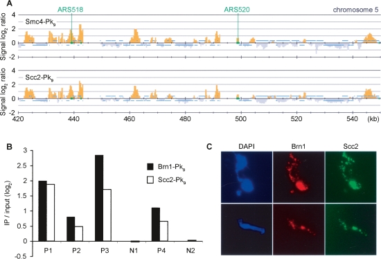
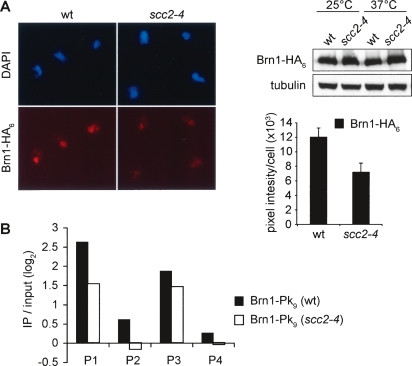
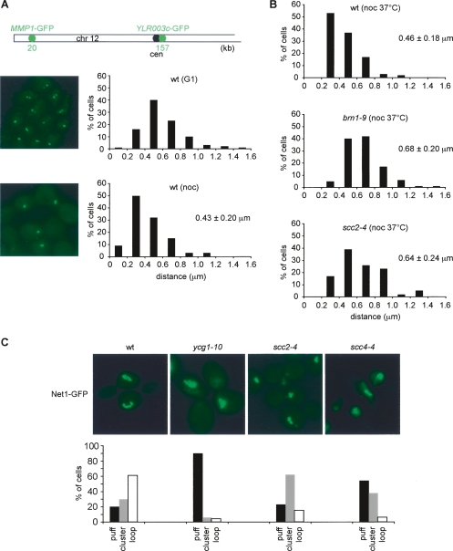
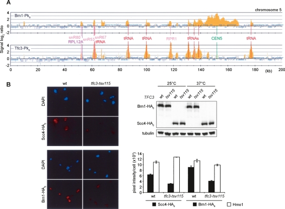
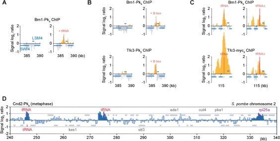
Eukaryotic chromosomes reach their stable rod-shaped appearance in mitosis in a reaction dependent on the evolutionarily conserved condensin complex. Little is known about how and where condensin associates with chromosomes. Here, we analyze condensin binding to budding yeast chromosomes using high-resolution oligonucleotide tiling arrays. Condensin-binding sites coincide with those of the loading factor Scc2/4 of the related cohesin complex. The sites map to tRNA and other genes bound by the RNA polymerase III transcription factor TFIIIC, and ribosomal protein and SNR genes. An ectopic B-box element, recognized by TFIIIC, constitutes a minimal condensin-binding site, and TFIIIC and the Scc2/4 complex promote functional condensin association with chromosomes. A similar pattern of condensin binding is conserved along fission yeast chromosomes. This reveals that TFIIIC-binding sites, including tRNA genes, constitute a hitherto unknown chromosomal feature with important implications for chromosome architecture during both interphase and mitosis.


Figures

Figure 1.
Figure 1.
A largely unaltered condensin pattern during the cell cycle. (A) Comparison of the condensin subunits Smc4-Pk9 and Brn1-Pk9 in G1 versus metaphase. Cultures of strain Y2315 (MATa SMC4-Pk9) and Y2200 (MATa BRN1-Pk9) were arrested in G1 by mating pheromone α-factor treatment, or in metaphase by addition of the spindle poison nocodazole. Uniform arrest was confirmed by flow cytometric analysis of DNA content. Enrichment of DNA fragments in condensin chromatin immunoprecipitates relative to a whole genome DNA sample is shown along a 325-kb region of budding yeast chromosome 5. Each bar represents the average of 16 oligonucleotide probes within adjacent 300-bp windows. Orange signals represent significant binding as described (Katou et al. 2003). Blue bars above and below the midline represent ORFs transcribed from left to right and opposite, respectively. The centromere and origins of replication are depicted in green. P1–P3 indicate three of the four positive, and N1 indicates one of the two negative, association sites that were confirmed by quantitative PCR (cf. Fig. 3). (B) Accumulation of Smc4 at stalled replication forks. Cells of strain Y2315 were synchronized in G1 by α-factor treatment and released into medium containing the replication inhibitor HU for 2 h. Cells of strain K5601 (MATa GPD-TK) were treated in the same way, but were released from α-factor arrest in the presence of 400 μg/mL bromodeoxyuridine (BrdU). ChIP against Smc4-Pk9 or BrdU was performed.
Figure 2.
Figure 2.
Condensin binding without a spacing rule. (A) Condensin binding sites are distinct from those of the related cohesin complex. The distribution of Smc4-Pk9 in nocodazole-arrested cells from the experiment in Figure 1 (significant peaks in blue) was overlaid with that of Scc1-HA6 (red) (Lengronne et al. 2004). The first 200 kb of chromosome 5 are shown. (B) Condensin binding does not follow a geometric spacing rule but adheres to sequence determinants. The pattern of Brn1-Pk9 is shown in strains Y2200 and Y2565 (MATa BRN1-Pk9 ChrVΔ213,898-222,402) harboring an 8.5-kb deletion between two neighboring condensin-binding sites.
Figure 3.
Figure 3.
Condensin colocalization with the cohesin loader Scc2/4. (A) Colocalization seen by ChIP. The Smc4-Pk9 pattern along a 130-kb fragment of chromosome 5 in HU-arrested cells is compared with the distribution of Scc2-Pk9 obtained from strain Y2422 (MATa SCC2-Pk9) under the same conditions. The locations of the early firing origins ARS518 and ARS520 are indicated. (B) Confirmation of colocalization by quantitative PCR analysis. Four positive (P1–P4) and two negative (N1 and N2) association sites identified by microarray analysis (cf. Fig. 1; Supplemental Fig. 1) were examined. Enrichment of Brn1-Pk9 (strain Y2200) and Scc2-Pk9 chromatin immunoprecipitates over a whole genome DNA sample is depicted, normalized to the average of the two negative association sites. 1.58% and 1.45% of input DNA at P1 was recovered in the Brn1 and Scc2 immunoprecipitates, respectively. (C) Colocalization on chromosome spreads. Strain Y2717 (MATa BRN1-myc9 SCC2-HA6) was arrested in metaphase by nocodazole treatment. Chromosome spreads were stained to detect the epitope-tagged condensin subunit Brn1-myc9 and Scc2-HA6.
Figure 4.
Figure 4.
A contribution of Scc2/4 to condensin association with chromosomes. (A) Chromosome spreads of nocodazole-arrested cells of strains Y2521 (MATa BRN1-HA6 SCC1-Pk9) and Y2423 (MATa scc2-4 BRN1-HA6 SCC1-Pk9) shifted for 1 h to 37°C were stained for the condensin subunit Brn1-HA6. The mean staining intensity and standard error for at least 50 cells in each sample are shown. The decrease of Brn1 on chromosomes was not due to degradation of the subunit, as confirmed by Western blotting of whole cell extracts. (B) Quantification of condensin association under the same conditions by ChIP followed by real-time PCR analysis of the four binding sites (P1–P4) characterized in Figure 3B. The two negative sites (N1 and N2) were used for normalization.
Figure 5.
Figure 5.
Scc2/4 requirement for chromosome condensation. (A) A condensation assay for the chromosome 12 left arm. Strain Y2869 (MATa lacOs∷YLR003c-1 lacOs∷MMP1 LacI-GFP) was arrested in G1 by α-factor, and in metaphase by nocodazole treatment. Chromosome condensation was assessed by measuring the distance between the two fluorescent loci in three dimensions. Examples of wild-type cells in G1 and metaphase are shown, as well as the distribution of measured distances. The mean distances, with their standard deviations, are given. (B) Scc2/4 is required to maintain chromosome arm condensation. Metaphase arrested cells of strains Y2869, Y3104 (as Y2869 but brn1-9) and Y2887 (as Y2869 but scc2-4) were shifted for 1 h to 35.5°C, and the condensation status of the chromosome 12 left arm was analyzed as in A. (C) Scc2/4 is required for condensation of the rDNA locus. Strains Y2727 (MATa NET1-GFP), Y2729 (MATα ycg1-10 NET1-GFP), Y2750 (MATα scc2-4 NET1-GFP), and Y2728 (MATα scc4Δ scc4-4 NET1-GFP) were arrested in S phase by HU treatment at 23°C, and released into nocodazole-containing medium at 35.5°C. rDNA condensation was analyzed by visualizing the rDNA-binding protein Net1 fused to GFP. Examples of the cells are shown, and the numbers of nuclei displaying puff-, cluster-, or loop-shaped rDNA loci (Lavoie et al. 2004) were counted in >100 cells in each sample.
Figure 6.
Figure 6.
Condensin coincides with the RNA pol III transcription factor TFIIIC. (A) Colocalization of condensin with TFIIIC at tRNA genes, and genes encoding ribosomal protein components and small noncoding RNAs. Strains Y2200 and Y3096 (MATα TFC3-Pk9 BRN1-HA6) were arrested in metaphase by nocodazole treatment, and ChIP against the condensin subunit Brn1-Pk9 and the TFIIIC subunit Tfc3-Pk9 was performed. A 200-kb region including the centromere of chromosome 5 is shown. (B) Reduced chromosome association of Scc2/4 and condensin in the temperature-sensitive TFIIIC mutant tfc3-tsv115. Cultures of strains Y3343 (MATa SCC4-HA6), Y3344 (as Y3343 but tfc3-tsv115), Y3345 (MATa BRN1-HA6 SCC1-Pk9), and Y3346 (as Y3345 but tfc3-tsv1) were shifted for 4 h to 37°C, and chromosome binding of Scc4-HA6 and Brn1-HA6 was quantified on stained spread chromosomes. Examples of the spreads and the measured mean staining intensity and standard error are shown.
Figure 7.
Figure 7.
A B-box as minimal condensin-binding site. (A) tRNA genes confer condensin binding. Brn1-Pk9 ChIP was performed in strains Y2200 and Y3528 [as Y2200, but two tandem tRNA tH(GUG)E1 genes inserted between SWI4 and LSM4] arrested in mitosis by nocodazole treatment. A 7.5-kb region around this locus is shown. All probes with a positive signal log2 ratio are depicted in yellow. (B) A B-box is sufficient to form a TFIIIC and condensin-binding site. As in A, but a B-box consensus sequence (GGTTCGAACCC) was inserted. Chromatin immunoprecipitates against Brn1-Pk9 (strains Y2200 and Y3485) or Tfc3-Pk9 (Y3096 and Y3475) were analyzed. (C) Additional determinants at an endogenous tRNA gene. Brn1-Pk9 and Tfc3-myc18 association around tRNA tE(UUC)J on chromosome 10 before (strains Y2200 and Y3096) and after deletion of the tRNA gene (Y3521 and Y3522) is shown. (D) Condensin association along fission yeast chromosome 2. Nda3-KM311 cells, Y253 (h- leu1 nda3-KM311 cnd2-Pk9), were arrested in metaphase by temperature shift for 6 h to 20°C, before ChIP against Cnd2-Pk9 was performed. The distribution over a 100-kb region of S. pombe chromosome 2 is shown. Peaks identified as detailed in Supplemental Figure 6 are indicated in dark blue.

References

    1. Anderson D.E., Losada A., Erickson H.P., Hirano T. Condensin and cohesin display different arm conformations with characteristic hinge angles. J. Cell Biol. 2002;156:419–424. - PMC - PubMed
    1. Aono N., Sutani T., Tomonaga T., Mochida S., Yanagida M. Cnd2 has dual roles in mitotic condensation and interphase. Nature. 2002;417:197–202. - PubMed
    1. Arumugam P., Gruber S., Tanaka K., Haering C.H., Mechtler K., Nasmyth K. ATP hydrolysis is required for cohesin’s association with chromosomes. Curr. Biol. 2003;13:1941–1953. - PubMed
    1. Bazett-Jones D.P., Kimura K., Hirano T. Efficient supercoiling of DNA by a single condensin complex as revealed by electron spectroscopic imaging. Mol. Cell. 2002;9:1183–1190. - PubMed
    1. Betts Lindroos H., Ström L., Itoh T., Katou Y., Shirahige K., Sjögren C. Chromosomal association of the Smc5/6 complex reveals that it functions in differently regulated pathways. Mol. Cell. 2006;22:755–767. - PubMed

Publication types

MeSH terms