. 2008 Aug 15;22(16):2267-77.
doi: 10.1101/gad.1670908.

Recognition of misfolded proteins by Lon, a AAA(+) protease

Affiliations

Recognition of misfolded proteins by Lon, a AAA(+) protease

Eyal Gur et al. Genes Dev. .

Abstract

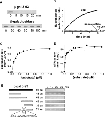
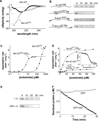
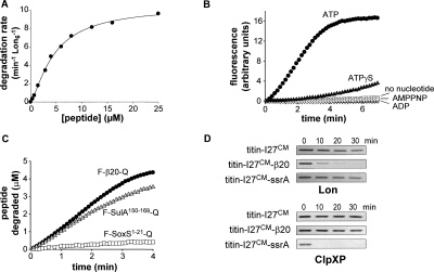
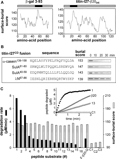
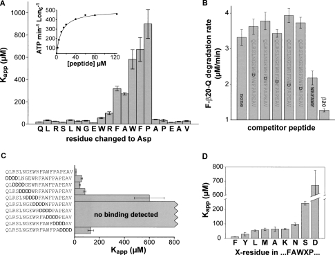
Proteins unfold constantly in cells, especially under stress conditions. Degradation of denatured polypeptides by Lon and related ATP-dependent AAA(+) proteases helps prevent toxic aggregates formation and other deleterious consequences, but how these destructive enzymatic machines distinguish between damaged and properly folded proteins is poorly understood. Here, we show that Escherichia coli Lon recognizes specific sequences -- rich in aromatic residues -- that are accessible in unfolded polypeptides but hidden in most native structures. Denatured polypeptides lacking such sequences are poor substrates. Lon also unfolds and degrades stably folded proteins with accessible recognition tags. Thus, protein architecture and the positioning of appropriate targeting sequences allow Lon degradation to be dependent or independent of the folding status of a protein. Our results suggest that Lon can recognize multiple signals in unfolded polypeptides synergistically, resulting in nanomolar binding and a mechanism for discriminating irreversibly damaged proteins from transiently unfolded elements of structure.


Figures

Figure 1.
Figure 1.
Lon recognition and degradation of denatured β-galactosidase fragments. (A) Lon (150 nM hexamer) degraded the 3–93 fragment of β-galactosidase (5 μM) but not native β-galactosidase (5 μM) as assayed by SDS-PAGE. (B) Rapid degradation of fluorescein-labeled 3–93 fragment (5 μM) by Lon (150 nM hexamer) required ATP and was not observed with ADP, without nucleotide, or without Lon. (C) Steady-state rates of degradation of different concentrations of the 3–93 fragment by Lon (10 nM hexamer) were assayed by release of acid soluble 14C-labeled peptides. The solid curve is a fit to the Henri-Michaelis-Menten equation (R2 = 0.99). KM and Vmax are listed in Table 1. (D) Stimulation of Lon S679A (10 nM hexamer) ATP-hydrolysis rates by increasing concentrations of the 3–93 fragment. The curve is a fit (R2 = 0.98) to the hyperbolic binding isotherm (rate = basal + max ⋅ [S]/(Kapp + [S])) with Kapp = 32 ± 3 nM. (E) Deletion analysis of the 3–93 fragment. Truncated variants of the 3–93 fragment (5 μM) were assayed for degradation by Lon (150 nM hexamer) by SDS-PAGE.
Figure 2.
Figure 2.
Properties and Lon degradation of substrates. (A) Circular–dichroism spectra (25°C) show that titin-I27 (40 μM) is natively folded and titin-I27CD (40 μM) is denatured. (B) SDS-PAGE assays of degradation of 5 μM titin-I27CD or β20-tagged variants by Lon (150 nM hexamer). The internal β20 tag was cloned between residues 17 and 18 of titin-I27CD. (C) Steady-state rates of Lon (100 nM hexamer) degradation of 35S-labeled titin-I27CD with or without an internal β20 insertion were assayed by release of acid-soluble peptides. The solid curves are fits (R2 = 0.99 for both curves) to the Hill equation (V = Vmax ⋅ [S]n/(KMn + [S]n); values for Vmax, KM, and the Hill constant are listed in Table 1. (D) Steady-state rates of degradation of 35S-labeled native titin-I27-β20 and denatured titin-I27CD-β20 by Lon (100 nM hexamer) were determined and fitted as described in C (R2 = 0.99 for both curves). Values of KM, Vmax, and the Hill constant are listed in Table 1. (Inset) As assayed by changes in circular–dichroism ellipticity at 228 nm, titin-I27 (closed symbols) and titin-I27-β20 (open symbols) had the same thermal stability. (E) SDS-PAGE assays of Lon degradation (150 nM hexamer) of the N-terminal domain of λ cI repressor (5 μM) with or without a β20 tag. (F) Degradation of GFP or GFP-β20 (3 μM each) by Lon (6 μM hexamer). Reactions were measured by decreases in GFP fluorescence.
Figure 3.
Figure 3.
Degradation directed by peptide-tag sequences (A) Concentration dependence of the steady-state rate of F-β20-Q degradation by Lon (100 nM hexamer). The solid line is a fit (R2 = 0.99) to the Hill equation (V = Vmax ⋅ [S]n/(KMn + [S]n). KM, Vmax, and Hill constants are listed in Table 1. (B) Degradation of the F-β20-Q peptide (5 μM) by Lon (0.3 μM hexamer) was assayed by increased fluorescence in the presence of 2 mM ATP, ATPγS, AMPPNP, or without nucleotide. No significant degradation was observed using LonS679A (0.3 μM hexamer) and ATP. (C) Degradation of F-β20-Q, F-SulA150–169-Q, and F-SoxS1–21-Q (5 μM each) by Lon (0.3 μM hexamer). (D) Degradation of unfolded titin substrates by Lon (0.3 μM hexamer) or ClpXP (0.3 μM ClpX6, 0.9 μM ClpP14) was assayed by SDS-PAGE.
Figure 4.
Figure 4.
Correlation of surface-burial scores with Lon degradation. (A) Surface-burial scores for β-gal 3–93 and titin-I27-β20int were calculated as described in Experimental Procedures. The β20 sequence scored highest in both substrates. (B) Sequences with the highest surface-burial scores from three Lon substrates were inserted by cloning between residues 17 and 18 of unfolded titin-I27CD and each fusion protein (5 μM) was assayed for degradation by Lon (150 nM hexamer) by SDS-PAGE. (C) Peptides corresponding to 15-residue β-galactosidase sequences with the highest surface-burial scores (numbered 1–16) were synthesized with flanking fluorophore and quencher groups. Each peptide (5 μM) was incubated with Lon (300 nM hexamer) and the rate of degradation was assayed by changes in fluorescence, as shown for representative peptides in the inset. C1–C3 are 15-residue control β-galactosidase peptides with low surface-burial scores. Degradation rates (dark bars) and surface-burial scores (light bars) are plotted for each peptide. Peptide sequences were RWQFNRQSGFLSQMW (1), YWQAFRQYP RLQGGF (2), FAKYWQAFRQYPRLQ (3), HYPNHPLWYTLC DRY (4), MWRMSGIFRDVSLLH (5), RWDLPLSDMYTPYVF (6), RWLPAMSERVTRMVQ (7), EYLFRHSDNELLHWM (8), YLEDQDMWRMSGIFR (9), LTEAKHQQQFFQFRL (10), LRA GENRLAVMVLRW (11), LLIRGVNRHEHHPLH (12), RMVQR DRNHPSVIIW (13), FRQYPRLQGGFVWDW (14), HQWRGD FQFNISRYS (15), FVWDWVDQSLIKYDE (16), GETQVASG TAPFGGE (C1), RPVQYEGGGADTTAT (C2), and GIGGDDS WSPSVSAE (C3).
Figure 5.
Figure 5.
Mutational analysis of β20-peptide recognition by Lon. (A) The apparent Lon affinities of β20-peptide variants, each containing a single aspartic acid substitution, were determined by assaying changes in the ATPase rate of Lon (150 nM hexamer) at a series of peptide concentrations (bars represent standard errors for the Kapp values). The inset shows the data and fitted affinity curve for the wild-type β20 peptide (Kapp = 10.7 ± 0.8 μM). R2 values for all fitted curves were >0.97. For weakly binding peptides, saturation was not reached and Κapp was calculated by assuming that Vmax was ≈500 ATP/min ⋅ Lon6. (B) Degradation of F-β20-Q (25 μM) by Lon (300 nM hexamer) was measured in the presence/absence of the designated competitor peptides (125 μΜ each). (R2 = 0.99 for both curves). (C) Values of Kapp for Lon binding were measured for β20-peptide variants in which a block of four consecutive residues was substituted with DDDD as described in A. R2 values for all fitted curves were >0.97. (D) Values of Kapp for Lon binding, measured as described in A, for β20-peptides with different residues at the X position in the FAWXP subsequence. Values for the F and D sequences are taken from A. R2 values for all fitted curves were >0.97.

References

    1. Baker T.A., Sauer R.T. ATP-dependent proteases of bacteria: Recognition logic and operating principles. Trends Biochem. Sci. 2006;31:647–653. - PMC - PubMed
    1. Baldwin E.P., Matthews B.W. Core-packing constraints, hydrophobicity and protein design. Curr. Opin. Biotechnol. 1994;5:396–402. - PubMed
    1. Bukau B., Weissman J., Horwich A. Molecular chaperones and protein quality control. Cell. 2006;125:443–451. - PubMed
    1. Burton R.E., Baker T.A., Sauer R.T. Nucleotide-dependent substrate recognition by the AAA+ HslUV protease. Nat. Struct. Mol. Biol. 2005;12:245–251. - PubMed
    1. Carrion-Vazquez M., Oberhauser A.F., Fowler S.B., Marszalek P.E., Broedel S.E., Clarke J., Fernandez J.M. Mechanical and chemical unfolding of a single protein: A comparison. Proc. Natl. Acad. Sci. 1999;96:3694–3699. - PMC - PubMed

Publication types

MeSH terms