

The volatile anesthetic isoflurane perturbs conformational activation of integrin LFA-1 by binding to the allosteric regulatory cavity
- PMID: 18708587
- PMCID: PMC2614612
- DOI: 10.1096/fj.08-113324
The volatile anesthetic isoflurane perturbs conformational activation of integrin LFA-1 by binding to the allosteric regulatory cavity
Abstract
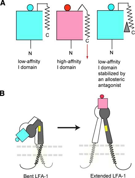
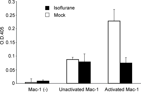
The molecular and structural basis of anesthetic interactions with conformations and functionalities of cell surface receptors remains to be elucidated. We have demonstrated that the widely used volatile anesthetic isoflurane blocks the activation-dependent conformational conversion of integrin lymphocyte function associated antigen-1 (LFA-1), the major leukocyte cell adhesion molecule, to a high-affinity configuration. Perturbation of LFA-1 activation by isoflurane at clinically relevant concentrations leads to the inhibition of T-cell interactions with target cells as well as ligand-triggered intracellular signaling. Nuclear magnetic resonance spectroscopy reveals that isoflurane binds within a cavity in the LFA-1 ligand-binding domain, which is a previously identified drug-binding site for allosteric small-molecule antagonists that stabilize LFA-1 in a low-affinity conformation. These results provide a potential mechanism for the immunomodulatory properties of isoflurane.
Figures
Similar articles
-
Sevoflurane binds and allosterically blocks integrin lymphocyte function-associated antigen-1.Anesthesiology. 2010 Sep;113(3):600-9. doi: 10.1097/ALN.0b013e3181e89a77. Anesthesiology. 2010. PMID: 20693879 Free PMC article.
-
Crystal structure of isoflurane bound to integrin LFA-1 supports a unified mechanism of volatile anesthetic action in the immune and central nervous systems.FASEB J. 2009 Aug;23(8):2735-40. doi: 10.1096/fj.09-129908. Epub 2009 Mar 30. FASEB J. 2009. PMID: 19332643 Free PMC article.
-
Small molecule inhibitors induce conformational changes in the I domain and the I-like domain of lymphocyte function-associated antigen-1. Molecular insights into integrin inhibition.J Biol Chem. 2002 Mar 22;277(12):10590-8. doi: 10.1074/jbc.M110521200. Epub 2002 Jan 7. J Biol Chem. 2002. PMID: 11781316
-
Lymphocyte function-associated antigen-1 blockade by statins: molecular basis and biological relevance.Endothelium. 2003;10(1):43-7. doi: 10.1080/10623320303360. Endothelium. 2003. PMID: 12699076 Review.
-
Integrin inside-out signaling and the immunological synapse.Curr Opin Cell Biol. 2012 Feb;24(1):107-15. doi: 10.1016/j.ceb.2011.10.004. Epub 2011 Nov 28. Curr Opin Cell Biol. 2012. PMID: 22129583 Free PMC article. Review.
Cited by
-
Sevoflurane binds and allosterically blocks integrin lymphocyte function-associated antigen-1.Anesthesiology. 2010 Sep;113(3):600-9. doi: 10.1097/ALN.0b013e3181e89a77. Anesthesiology. 2010. PMID: 20693879 Free PMC article.
-
Volatile Anesthetic Sevoflurane Attenuates Toll-Like Receptor 1/2 Activation.Anesth Analg. 2020 Aug;131(2):631-639. doi: 10.1213/ANE.0000000000004741. Anesth Analg. 2020. PMID: 32149756 Free PMC article.
-
Sedative drug modulates T-cell and lymphocyte function-associated antigen-1 function.Anesth Analg. 2011 Apr;112(4):830-8. doi: 10.1213/ANE.0b013e31820dcabb. Epub 2011 Mar 8. Anesth Analg. 2011. PMID: 21385989 Free PMC article.
-
Propofol shares the binding site with isoflurane and sevoflurane on leukocyte function-associated antigen-1.Anesth Analg. 2013 Oct;117(4):803-811. doi: 10.1213/ANE.0b013e3182a00ae0. Epub 2013 Aug 19. Anesth Analg. 2013. PMID: 23960033 Free PMC article.
-
Volatile anesthetics affect macrophage phagocytosis.PLoS One. 2019 May 9;14(5):e0216163. doi: 10.1371/journal.pone.0216163. eCollection 2019. PLoS One. 2019. PMID: 31071106 Free PMC article.
References
-
- McBride W T, Armstrong M A, McBride S J. Immunomodulation: an important concept in modern anaesthesia. Anaesthesia. 1996;51:465–473. - PubMed
-
- Heindl B, Reichle F M, Zahler S, Conzen P F, Becker B F. Sevoflurane and isoflurane protect the reperfused guinea pig heart by reducing postischemic adhesion of polymorphonuclear neutrophils. Anesthesiology. 1999;91:521–530. - PubMed
-
- Mobert J, Zahler S, Becker B F, Conzen P F. Inhibition of neutrophil activation by volatile anesthetics decreases adhesion to cultured human endothelial cells. Anesthesiology. 1999;90:1372–1381. - PubMed
-
- Hayes J K, Havaleshko D M, Plachinta R V, Rich G F. Isoflurane pretreatment supports hemodynamics and leukocyte rolling velocities in rat mesentery during lipopolysaccharide-induced inflammation. Anesth Analg. 2004;98:999–1006. - PubMed
Publication types
MeSH terms
Substances
Grants and funding
LinkOut - more resources
Full Text Sources
Other Literature Sources
