. 2008 Dec;22(12):4218-27.
doi: 10.1096/fj.08-113076. Epub 2008 Aug 15.

NFAT regulates induction of COX-2 and apoptosis of keratinocytes in response to ultraviolet radiation exposure

Affiliations

NFAT regulates induction of COX-2 and apoptosis of keratinocytes in response to ultraviolet radiation exposure

R J Flockhart et al. FASEB J. 2008 Dec.

Abstract

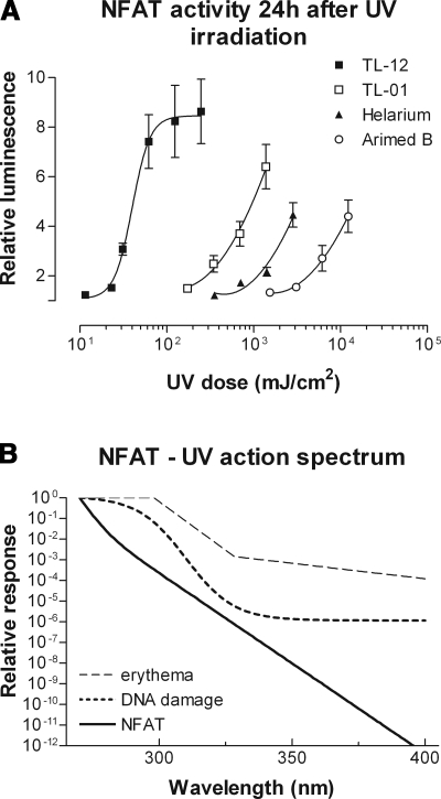
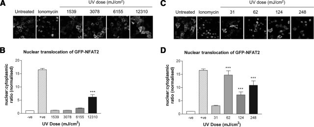
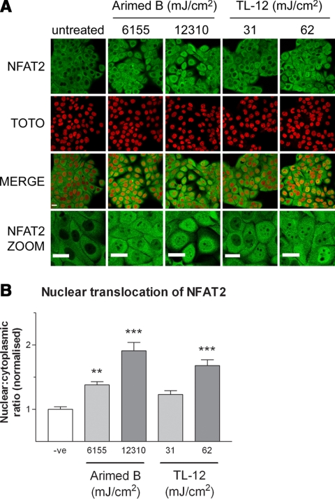
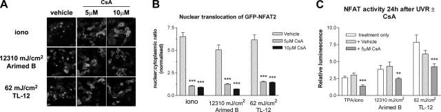
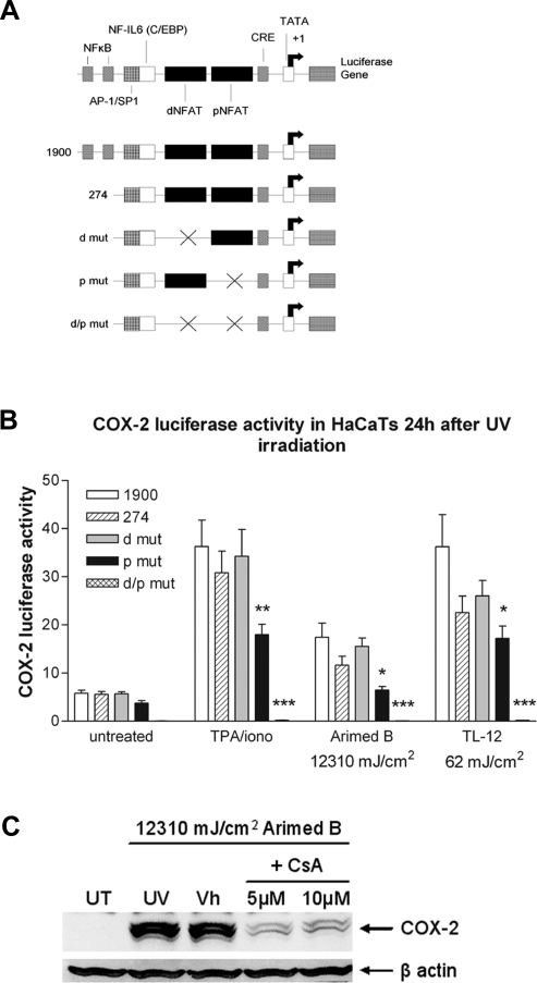
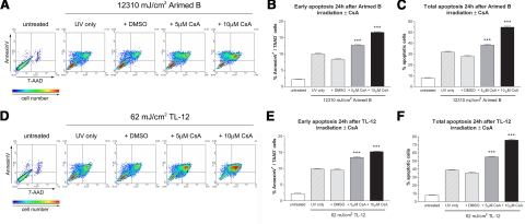
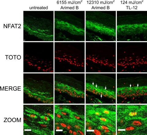
The nuclear factor of activated T cells (NFAT) transcription factors are regulated by calcium/calcineurin signals and play important roles in T cells, muscle, bone, and neural tissue. NFAT is expressed in the epidermis, and although recent data suggest that NFAT is involved in the skin's responses to ultraviolet radiation (UVR), the wavelengths of radiation that activate NFAT and the biological function of UV-activated NFAT remain undefined. We demonstrate that NFAT transcriptional activity is preferentially induced by UVB wavelengths in keratinocytes. The derived action spectrum for NFAT activation indicates that NFAT transcriptional activity is inversely associated with wavelength. UVR also evoked NFAT2 nuclear translocation in a parallel wavelength-dependent fashion and both transcriptional activation and nuclear translocation were inhibited by the calcineurin inhibitor cyclosporin A. UVR also evoked NFAT2 nuclear translocation in three-dimensional skin equivalents. Evidence suggests that COX-2 contributes to UV-induced carcinogenesis. Inhibiting UV-induced NFAT activation in keratinocytes, reduced COX-2 protein induction, and increased UV-induced apoptosis. COX-2 luciferase reporters lacking functional NFAT binding sites were less responsive to UVR, highlighting that NFAT is required for UV-induced COX-2 induction. Taken together, these data suggest that the proinflammatory, antiapoptotic, and procarcinogenic functions of UV-activated COX-2 may be mediated, in part, by upstream NFAT signaling.


Figures

Figure 1.
Figure 1.
Ultraviolet radiation increases NFAT transcriptional activity: derivation of the NFAT action spectrum. A) NFAT luciferase activity was measured 24 h after UV irradiation with one of four different UV sources. All UV sources evoked a dose-dependent increase in NFAT transcriptional activity, with TL-12 lamps (53% UVB) being most effective. Values are means ± se from 3 independent experiments performed in triplicate. B) The action spectrum for NFAT was derived from the above luciferase data by a process of mathematical induction exploring three candidate models (see Materials and Methods). NFAT activity is inversely associated with wavelength. The action spectra for erythema and general DNA damage are shown for reference.
Figure 2.
Figure 2.
UVR evokes wavelength-dependent nuclear translocation of GFP-NFAT2. Cells were retrovirally transduced with virus transmitting GFP-NFAT2 prior to irradiation. A, C) Images show nuclear translocation 24 h after irradiation. Arimed B irradiation (A) (≥12,310 mJ/cm2) and TL-12 irradiation (C) (≥62 mJ/cm2) evoke nuclear translocation of GFP-NFAT2. B, D) Quantification of nuclear translocation by calculating nuclear:cytoplasmic ratio of GFP-NFAT2 following Arimed B irradiation (B) and TL-12 irradiation (D). Values are means ± se from at least 12 fields of view (≥90 cells in total) obtained from 3 independent experiments. Negative controls (–ve) were mock irradiated, and positive controls (+ve) were treated with 1 μM ionomycin. ***P < 0.001 vs. negative control; one-way ANOVA. Scale bar = 50 μm.
Figure 3.
Figure 3.
UVR evokes wavelength-dependent nuclear translocation of endogenous NFAT2. Cells were fixed 24 h after UV irradiation and immunostained for detection of endogenous NFAT2. A) Images represent the effect of UVR on endogenous NFAT2 localization 24 h after irradiation with Arimed B or TL-12 lamps. Digital zoom images are also included for clarity. B) Quantification of nuclear translocation by calculating nuclear:cytoplasmic ratio of NFAT2. Values are means ± se from 8 fields of view (≥161 cells in total) obtained from 3 independent experiments. Negative controls (–ve) were mock irradiated. **P < 0.01, ***P < 0.001 vs. negative control; one-way ANOVA. Scale bars = 50 μm.
Figure 4.
Figure 4.
CsA inhibits UV-induced GFP-NFAT2 nuclear translocation and NFAT transcriptional activation. A) Cells retrovirally transduced with virus transmitting GFP-NFAT2 were treated with either vehicle or 5 or 10 μM CsA for 16 h prior to UV irradiation or ionomycin (1 μM) treatment. Images show nuclear translocation 24 h after irradiation or ionomycin treatment. B) Quantification of nuclear translocation by calculating nuclear:cytoplasmic ratio of GFP-NFAT2. Values are means ± se from 8 fields of view (≥90 cells in total) obtained from 3 independent experiments. C) Cells were treated with either vehicle or 5 μM CsA 16 h prior to UV irradiation or TPA/ionomycin (TPA/iono) treatment, and NFAT luciferase activity was measured 24 h later. Values are means ± se from 3 independent experiments performed in triplicate. **P < 0.01, ***P < 0.001 vs. vehicle control; one-way ANOVA.
Figure 5.
Figure 5.
NFAT is involved in UV-induced activation of the COX-2 promoter and UV-induced COX-2 protein production. A) Schematic diagram of the human COX-2 promoter and the promoters of COX-2 luciferase reporters used (adapted from ref. 42). B) COX-2 promoter activity 24 h after UV irradiation was measured by transfecting cells with luciferase reporter vectors that selectively lacked either one or both of the NFAT binding sites present in the COX-2 promoter. Positive controls (TPA/iono) were treated with TPA (50 nM) and ionomycin (1 μM). Values are means ± se from 3 independent experiments performed in quadruplicate. C) Western blot analysis of lysates prepared 24 h after Arimed B irradiation in the presence or absence of 5 or 10 μM CsA pretreatment. The two COX-2 bands migrate to 70 and 72 kDa, which are glycosylated variants of COX-2 protein that have been previously described . *P < 0.05, ***P < 0.001 vs. control vector containing all NFAT binding sites (hatched bar); one-way ANOVA. UT, untreated; UV, UVR only; Vh, vehicle + UVR.
Figure 6.
Figure 6.
Inhibiting NFAT with CsA increases apoptosis in response to UV irradiation. Cells were treated with 5 or 10 μM CsA prior to irradiation. Density plots show that CsA pretreatment dose dependently increased UV-induced apoptosis after both Arimed B (A) and TL-12 (D) irradiation. The percentage of Annexin V positive/7AAD negative cells (early apoptosis) following Arimed B (B) and TL-12 (E) irradiation and percentage of Annexin V positive/7AAD positive cells plus Annexin V positive/7AAD negative cells (total apoptosis) following Arimed B (C), and TL-12 (F) irradiation is summarized in bar charts that show the mean ± se percentage of positive cells from 3 independent, triplicate experiments. ***P < 0.001 vs. vehicle (DMSO) control; one-way ANOVA.
Figure 7.
Figure 7.
UVR evokes NFAT2 nuclear translocation in a skin equivalent model. Full-thickness epidermal equivalents were irradiated with 6155 and 12,310 mJ/cm2 using Arimed B lamps or 124 mJ/cm2 using TL-12 lamps. Specimens were fixed 24 h after irradiation, and immunostaining for detection of endogenous NFAT2 was performed. Arrows indicate positive nuclear staining of NFAT2. Scale bars = 50 μm.

Similar articles

Cited by

References

    1. Shaw J P, Utz P J, Durand D B, Toole J J, Emmel E A, Crabtree G R. Identification of a putative regulator of early T cell activation genes. Science. 1988;241:202–205. - PubMed
    1. Kiani A, Rao A, Aramburu J. Manipulating immune responses with immunosuppressive agents that target NFAT. Immunity. 2000;12:359–372. - PubMed
    1. Hill-Eubanks D C, Gomez M F, Stevenson A S, Nelson M T. NFAT regulation in smooth muscle. Trends Cardiovasc Med. 2003;13:56–62. - PubMed
    1. Schulz R A, Yutzey K E. Calcineurin signaling and NFAT activation in cardiovascular and skeletal muscle development. Dev Biol. 2004;266:1–16. - PubMed
    1. Asagiri M, Sato K, Usami T, Ochi S, Nishina H, Yoshida H, Morita I, Wagner E F, Mak T W, Serfling E, Takayanagi H. Autoamplification of NFATc1 expression determines its essential role in bone homeostasis. J Exp Med. 2005;202:1261–1269. - PMC - PubMed

Publication types

MeSH terms