. 2008 Nov;295(5):L733-43.
doi: 10.1152/ajplung.90240.2008. Epub 2008 Aug 15.

Mitigation of chlorine-induced lung injury by low-molecular-weight antioxidants

Affiliations

Mitigation of chlorine-induced lung injury by low-molecular-weight antioxidants

Martin Leustik et al. Am J Physiol Lung Cell Mol Physiol. 2008 Nov.

Abstract

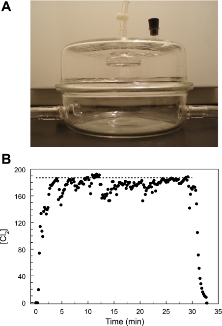
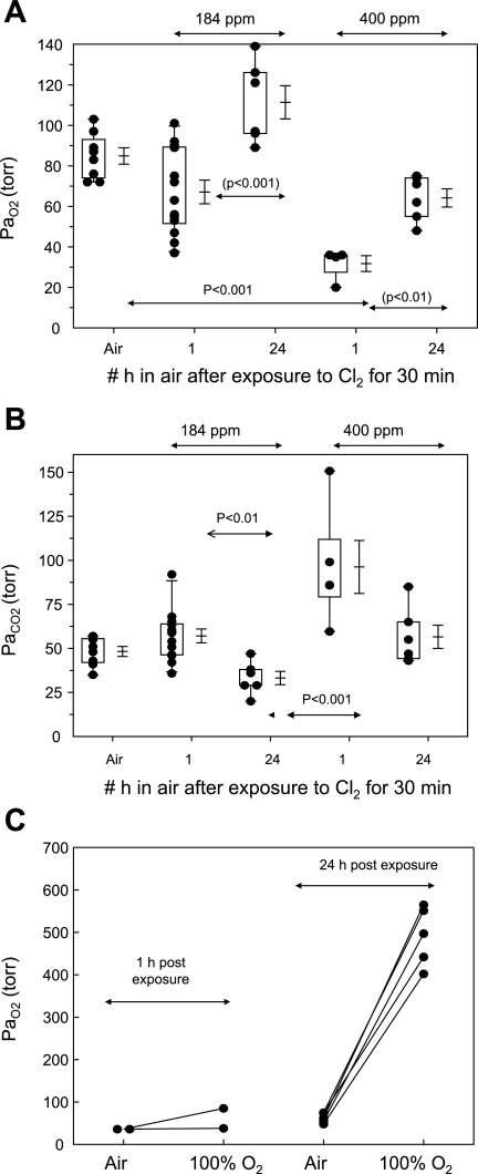
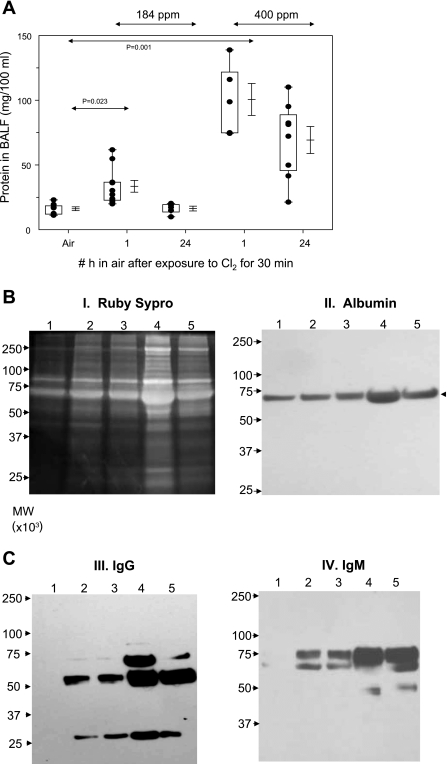
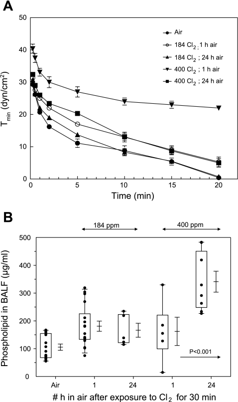
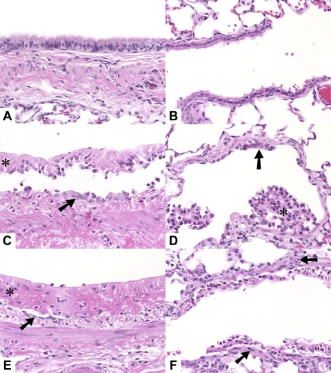
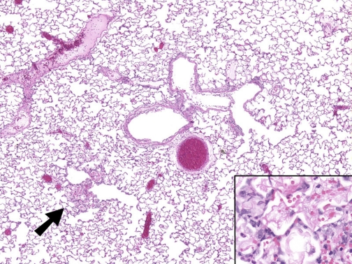
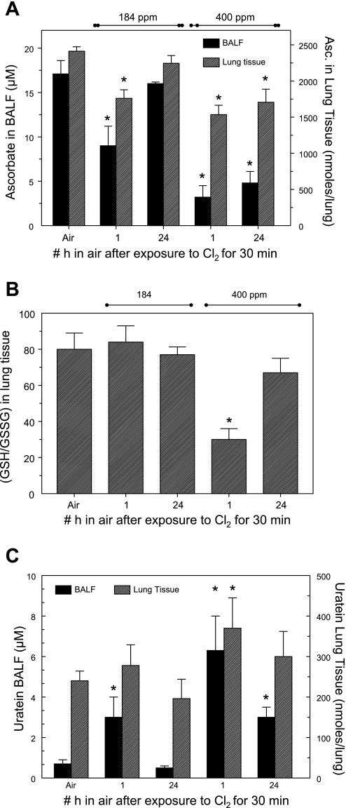
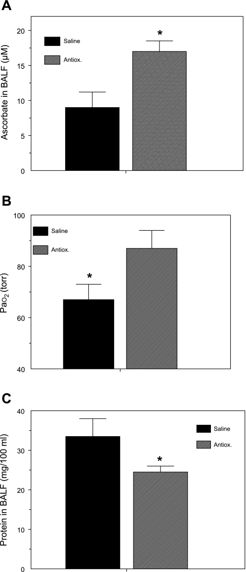
Chlorine (Cl(2)) is a highly reactive oxidant gas used extensively in a number of industrial processes. Exposure to high concentrations of Cl(2) results in acute lung injury that may either resolve spontaneously or progress to acute respiratory failure. Presently, the pathophysiological sequelae associated with Cl(2)-induced acute lung injury in conscious animals, as well as the cellular and biochemical mechanisms involved, have not been elucidated. We exposed conscious Sprague-Dawley rats to Cl(2) gas (184 or 400 ppm) for 30 min in environmental chambers and then returned them to room air. At 1 h after exposure, rats showed evidence of arterial hypoxemia, respiratory acidosis, increased levels of albumin, IgG, and IgM in bronchoalveolar lavage fluid (BALF), increased BALF surfactant surface tension, and significant histological injury to airway and alveolar epithelia. These changes were more pronounced in the 400-ppm-exposed rats. Concomitant decreases of ascorbate (AA) and reduced glutathione (GSH) were also detected in both BALF and lung tissues. In contrast, heart tissue AA and GSH content remained unchanged. These abnormalities persisted 24 h after exposure in rats exposed to 400 ppm Cl(2). Rats injected systemically with a mixture of AA, deferoxamine, and N-acetyl-L-cysteine before exposure to 184 ppm Cl(2) had normal levels of AA, lower levels of BALF albumin and normal arterial Po(2) and Pco(2) values. These findings suggest that Cl(2) inhalation damages both airway and alveolar epithelial tissues and that resulting effects were ameliorated by prophylactic administration of low-molecular-weight antioxidants.


Figures

Fig. 1.
Fig. 1.
A: glass chamber used for exposing rats to either Cl2 or air (see materials and methods for details). B: record of Cl2 concentration ([Cl2]) in the exposure chamber with 2 rats present. Rats were placed in the chamber and breathed room air for ∼10 min. At time 0, one of the mass flow controllers was connected to a Cl2 cylinder (1,000 ppm in air) while the other one remained connected to room air. Relative flow rates were adjusted to achieve a nominal concentration of 184 ppm (shown by dotted line; total flow rate 5 l/min). [Cl2] was monitored continuously every 2 s, and the output was stored in a portable computer (as shown by individual symbols). At 30 min the Cl2 cylinder was switched off and the compressed air flow rate was increased to 5 l/min. Measured [Cl2] was lower than the nominal value because of the absorption of Cl2 by the rats.
Fig. 2.
Fig. 2.
Arterial Po2 (PaO2) of air- and Cl2-exposed rats. Rats were exposed to either 184 or 400 ppm Cl2, after which they resumed breathing room air. They were anesthetized after 1 or 24 h postexposure, and an arterial blood sample was obtained from the right carotid artery while they breathed air for the measurement of PaO2 (A) and arterial Pco2 (PaCO2; B). Box-whisker plots show individual points (each dot is a different rat), boxes (25–75% percentile of the data), whiskers (lower 5th and upper 95% percentile of the data) as well as means ± SE. Statistical analysis of mean values was performed by 1-way analysis of variance followed by the Bonferroni modification of the t-test. C: PaO2 values of rats exposed to 400 ppm Cl2 and breathing room air for either 1 or 24 h. PaO2 values were measured in arterial blood samples while rats breathed air or 100% O2 for 15 min. Each point represents a rat.
Fig. 3.
Fig. 3.
Protein values in bronchoalveolar lavage fluid (BALF) after Cl2 exposure. Rats were exposed to either 184 or 400 ppm Cl2, after which they resumed breathing room air. They were killed 1 or 24 h after exposure, and their lungs were lavaged as described in materials and methods. A: protein values in BALF. Box-whisker plot showing individual points (each dot is a different rat), boxes (25–75% percentile of the data), whiskers (lower 5th and upper 95% percentile of the data), as well as means ± SE. Statistical analysis of mean values was performed by 1-way analysis of variance followed by the Bonferroni modification of the t-test. B and C: equal volumes of BALF proteins (40 μl) were separated by SDS-PAGE (10%) and visualized with Sypro Ruby staining (I) or transferred to nitrocellulose and immunoblotted for albumin [molecular weight (MW) ∼69,000; II], IgG (MW ∼55,000 for heavy chain and ∼25,000 for light chain; III), and IgM (MW ∼75,000; IV). Lane 1, control (air); lanes 2 and 3, 184 ppm Cl2 for 30 min followed by 1 or 24 h in air; lanes 4 and 5, 400 ppm Cl2 for 30 min followed by 1 or 24 h in air. Typical records shown were repeated 2 times with BALF from different rats with identical results.
Fig. 4.
Fig. 4.
Surfactant minimum surface tension and BALF phospholipid levels. A: minimum surface tension [Tmin (dyn/cm2)] of cell-free BALF, reconstituted at 1 mg/ml, measured in a bubble surfactometer during dynamic compression of a bubble to 50% of its original area as described in materials and methods. x-Axis shows the accumulative time that the bubble was oscillated. Values are means ± SE; n = 3 for each value. Note very high values of Tmin in BALF of rats exposed to 400 ppm Cl2 and returned to room air for 1 h. Values of BALF from 400 ppm Cl2-exposed rats as well as 184 ppm Cl2 and 1 h in air were significantly different from the corresponding air values (with t-test; P < 0.05). B: phospholipids in BALF: total phospholipid levels in BALF, pelleted by centrifugation at 12,500 g as described in the text. Box-whisker plot shows individual points (each dot is a different rat), boxes (25–75% percentile of the data), whiskers (5th and 95th percentiles of the data), as well as means ± SE.
Fig. 5.
Fig. 5.
Hematoxylin and eosin (H & E) sections of lungs of rats exposed to either air or Cl2: control (A and B), 1 h after exposure to 400 ppm Cl2 for 30 min (C and D), and 24 h after exposure to 400 ppm Cl2 for 30 min (E and F). A, C, and E: main stem bronchus. B, D, and F: terminal bronchiole and adjacent alveoli. C, D, and E: necrotic epithelium (*), flattened remnant epithelial cells (arrow). F: necrotic epithelium, sparse neutrophil accumulation in wall (arrows). Original magnification ×40, H & E stain.
Fig. 6.
Fig. 6.
H & E section of lung of rat exposed to Cl2. Mild focal alveolitis (arrow) characterized by alveolar hemorrhage and fibrin and sparse neutrophils (inset). Original magnifications ×4 and ×40 (inset), H & E stain.
Fig. 7.
Fig. 7.
Antioxidant levels in BALF and lung tissue of Cl2-exposed rats. A: ascorbate in cell-free BALF (left y-axis) and lung tissue (right y-axis) of air- and Cl2-exposed rats measured by HPLC as described in materials and methods. Values are means ± SE; no. of rats for BALF and lung tissue in each group: air, 9 and 7; 184 ppm Cl2 and 1 h in air, 12 and 11; 184 ppm Cl2 and 24 h in air, 4 and 5; 400 ppm Cl2 and 1 h in air, 6 and 7; 400 ppm Cl2 and 24 h in air, 8 and 8. *P < 0.05 vs. corresponding control (air-only value). B: reduced glutathione (GSH)-to-oxidized glutathione (GSSG) ratio values in lung of Cl2-exposed rats. Values are means ± SE; no. of rats in each group: air, 10; 184 ppm Cl2 and 1 h in air, 11; 184 ppm Cl2 and 24 h in air, 5; 400 ppm Cl2 and 1 h in air, 7; 400 ppm Cl2 and 24 h in air, 8. *P < 0.05 vs. corresponding control (air-only value). C: urate in cell-free BALF (left y-axis) and lung tissue (right y-axis) of air- and Cl2-exposed measured by HPLC as described in materials and methods. Values are means ± SE; no. of rats for BALF and lung tissue in each group: Air, 9 and 7; 184 ppm Cl2 and 1 h in air, 12 and 11; 184 ppm Cl2 and 24 h in air, 4 and 5; 400 ppm Cl2 and 1 h in air, 6 and 7; 400 ppm Cl2 and 24 h in air, 8 and 8. *P < 0.05 vs. corresponding control (air-only value).
Fig. 8.
Fig. 8.
Systemic administration of antioxidants decreases lung injury. Rats were injected with a mixture of antioxidants [Antiox; ascorbate, N-acetyl-l-cysteine (NAC), and deferoxamine] or an equivalent amount of vehicle (saline) 18 h and 1 h before being exposed to 184 ppm Cl2 for 30 min, returned to room air, and killed 1 h later, as described in materials and methods. Values are means ± SE. A: BALF ascorbate; no. of rats: saline, 5; Antiox, 16. B: PaO2 (from samples from carotid artery drawn during anesthesia); no. of rats: saline, 13; Antiox, 13. C: BALF protein; no. of rats: saline, 10; Antiox, 15; *P < 0.05 vs. corresponding saline values.

References

    1. Atkuri KR, Mantovani JJ, Herzenberg LA, Herzenberg LA. N-acetylcysteine—a safe antidote for cysteine/glutathione deficiency. Curr Opin Pharmacol 7: 355–359, 2007. - PMC - PubMed
    1. Ballinger CA, Cueto R, Squadrito G, Coffin JF, Velsor LW, Pryor WA, Postlethwait EM. Antioxidant-mediated augmentation of ozone-induced membrane oxidation. Free Radic Biol Med 38: 515–526, 2005. - PubMed
    1. Batchinsky AI, Martini DK, Jordan BS, Dick EJ, Fudge J, Baird CA, Hardin DE, Cancio LC. Acute respiratory distress syndrome secondary to inhalation of chlorine gas in sheep. J Trauma 60: 944–956, 2006. - PubMed
    1. Beckman JS, Beckman TW, Chen J, Marshall PA, Freeman BA. Apparent hydroxyl radical production by peroxynitrite: implications for endothelial injury from nitric oxide and superoxide. Proc Natl Acad Sci USA 87: 1620–1624, 1990. - PMC - PubMed
    1. Bell DG Management of acute respiratory distress syndrome (ARDS) following chlorine exposure (Abstract). Am J Respir Crit Care Med 176: A314, 2008.

Publication types

MeSH terms

LinkOut - more resources