Review
. 2008:42:517-40.
doi: 10.1146/annurev.genet.42.110807.091432.

Planar cell polarity signaling: from fly development to human disease

Affiliations
Review

Planar cell polarity signaling: from fly development to human disease

Matias Simons et al. Annu Rev Genet. 2008.

Abstract

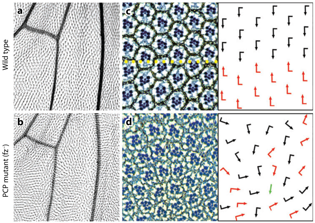
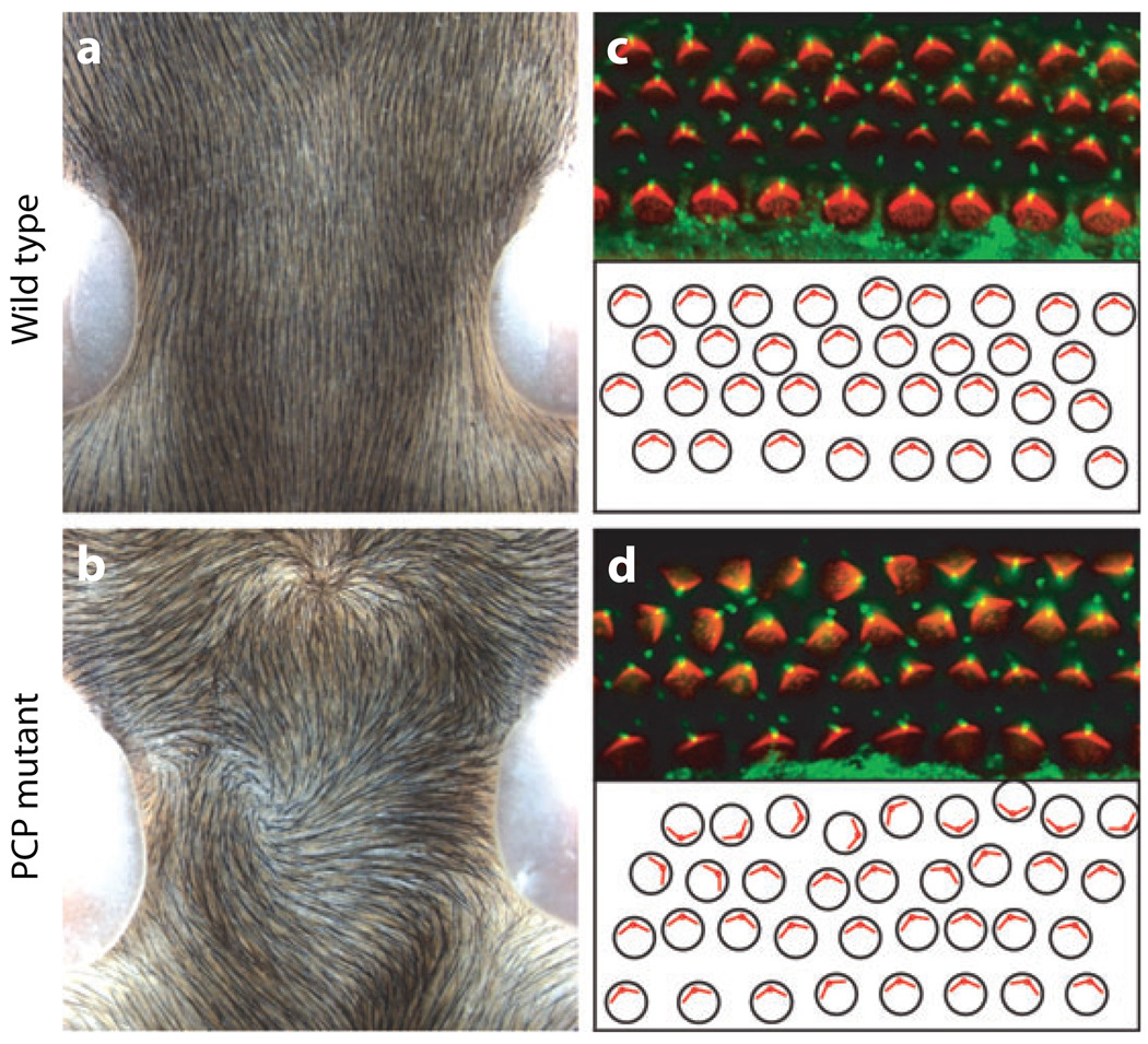
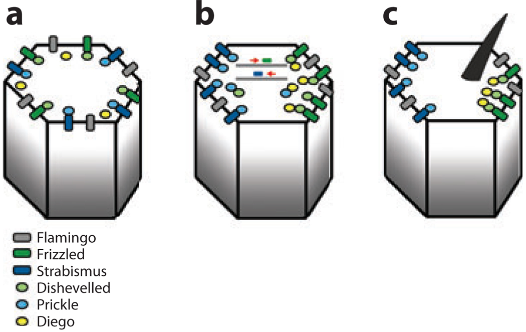
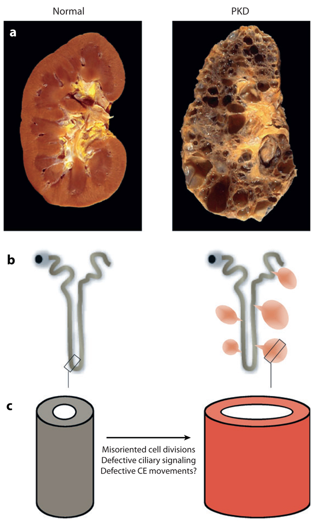
Most, if not all, cell types and tissues display several aspects of polarization. In addition to the ubiquitous epithelial cell polarity along the apical-basolateral axis, many epithelial tissues and organs are also polarized within the plane of the epithelium. This is generally referred to as planar cell polarity (PCP; or historically, tissue polarity). Genetic screens in Drosophila pioneered the discovery of core PCP factors, and subsequent work in vertebrates has established that the respective pathways are evolutionarily conserved. PCP is not restricted only to epithelial tissues but is also found in mesenchymal cells, where it can regulate cell migration and cell intercalation. Moreover, particularly in vertebrates, the conserved core PCP signaling factors have recently been found to be associated with the orientation or formation of cilia. This review discusses new developments in the molecular understanding of PCP establishment in Drosophila and vertebrates; these developments are integrated with new evidence that links PCP signaling to human disease.


Figures

Figure 1
Figure 1
Typical examples of PCP features in Drosophila. PCP effects in the wing (a,b) and eye (c,d). Wild type is shown in panels a and c (note the regular arrangements in both tissues), and the mutant appearance (fz) of same tissues is shown in b and d. (c and d) The right panels show the orientation of ommatidia schematically by arrows: black and red arrows represent the dorsal and ventral orientations (not mirror image symmetry in wild type/c) and loss of it in the mutant scenario. Occasional symmetrical ommatida (green arrow) are also found in the mutants.
Figure 2
Figure 2
Examples of PCP features in mammals. PCP features of the mouse skin (a,b) and the inner ear (c,d). (a,b) Dorsal view of mouse neck and the orientation of the skin hair in wild-type mice (a) and fz3 mutant mice (b). Note random whorls, swirls, and waves in the mutant genotype as compared to the normal anterior-posterior orientation in a. In a and b anterior is up. (c,d) Orientation of sensory hair cells of the mammalian (mouse) chochlea (inner ear). Each cell contains polarized bundles of actin-based stereocilia (labeled red with phalloidin) and a tubulin-based kinocilium (labeled with antiacetylated tubulin in green). In PCP mutants these bundles still form but their orientation becomes randomized [d; Looptail/Vangl2 (stbm) mutant]. The lower panels in c and d show schematic representation of the cellular (actin bundle) orientation reflecting their randomized appearance in the mutant. The original pictures were kindly provided by Jeremy Nathans.
Figure 3
Figure 3
Schematic presentation of the generation of asymmetric core PCP protein localization in Drosophila wing cells. In pupal wing cells, the core PCP proteins of the Fz/Fmi cassette become asymmetrically localized to proximal and distal cell membranes. Proximal is left and distal is right in all panels. A single cell at different stages maturing from left to right is shown in the respective panels ac. (a) Schematic of the localization of the core PCP proteins prior to any asymmetry detection at the onset of their interactions. (b) During polarization the Fz-Dsh-Dgo complexes become enriched at the distal end of each cell, whereas the Stbm-Pk complexes concentrate proximally. Fz has been seen to travel distally on microtubule-associated particles (121), and something similar might also happen for Stbm/Vang. (c) Final stage of polarization with all complexes resolved in either distal (Fz-associated protein or proximal cell ends. An actin-based hair is formed close to the distal vertex of each cell (where Fz-Dsh are localized). The Drosophila pupal wing is the only tissue in which the localization of all core PCP proteins has been analyzed; all but Dsh have also been analyzed in the Drosophila eye.
Figure 4
Figure 4
PCP and polycystic kidney disease (a) Photographs of a healthy (left) and a polycystic kidney (right). (b) Schematic diagram of the nephron, the functional unit of the kidney. In ADPKD, cysts arise from all segments of the nephron (right panel). (c) Cyst formation is thought to be caused, in part, by defective PCP signaling. The PCP pathway regulates the cell division plane of epithelial cells in the elongating tube during renal development. More speculatively, elongation of the growing tube could also be driven by convergent extension-like movements. The cilium might provide directional cues for the regulation of these two processes.

References

    1. Adler PN. Planar signaling and morphogenesis in Drosophila. Dev. Cell. 2002;2:525–535. - PubMed
    1. Adler PN, Liu J, Charlton J. Cell size and the morphogenesis of wing hairs in Drosophila. Genesis. 2000;28:82–91. - PubMed
    1. Ahumada A, Slusarski DC, Liu X, Moon RT, Malbon CC, Wang HY. Signaling of rat Frizzled-2 through phosphodiesterase and cyclic GMP. Science. 2002;298:2006–2010. - PubMed
    1. Axelrod JD. Basal bodies, kinocilia and planar cell polarity. Nat. Genet. 2008;40:10–11. - PubMed
    1. Axelrod JD, Miller JR, Shulman JM, Moon RT, Perrimon N. Differential requirement of Dishevelled provides signaling specificity in the Wingless and planar cell polarity signaling pathways. Genes Dev. 1998;12:2610–2622. - PMC - PubMed

Publication types

MeSH terms

LinkOut - more resources