

Interphase cytogenetics for 1p19q and t(1;19)(q10;p10) may distinguish prognostically relevant subgroups in extraventricular neurocytoma
- PMID: 18710393
- PMCID: PMC2742575
- DOI: 10.1111/j.1750-3639.2008.00206.x
Interphase cytogenetics for 1p19q and t(1;19)(q10;p10) may distinguish prognostically relevant subgroups in extraventricular neurocytoma
Abstract
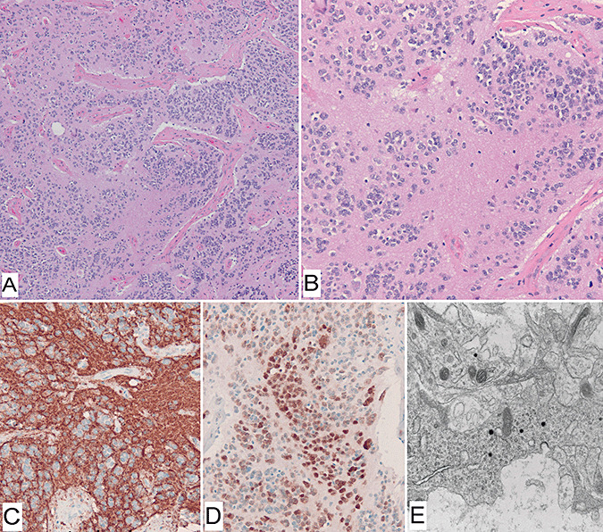
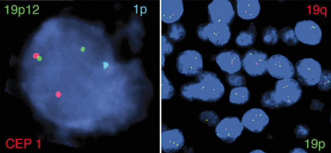
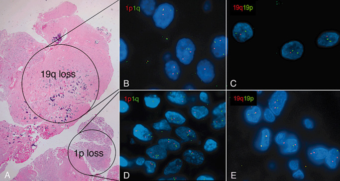
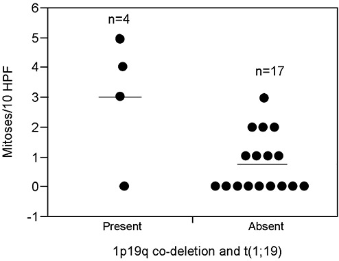
Co-deletion of chromosome arms 1p and 19q, characteristic of oligodendroglial tumors, was recently found to be mediated by t(1;19)(q10;p10). To evaluate the prevalence of 1p19q co-deletion and t(1;19) in extraventricular neurocytomas (EVN), we studied tumors from 23 patients, including 13 females and 10 males (median age at diagnosis 34 years, range 2-76 years). Fluorescence in situ hybridization (FISH) studies were performed with probes targeting 1p36/1q25 and 19q13/19p13 to assess for 1p19q co-deletion, as well as chromosome 1 alpha-satellite and 19p12 to detect t(1;19)(q10;p10). FISH was successful in 21 (91%) cases and demonstrated 1p19q co-deletion in five cases (24%) or isolated 1p loss in two cases (10%). Evidence for t(1;19) was found in four (of five) cases with 1p19q co-deletion. Three tumors with 1p19q loss and t(1;19) demonstrated atypical histologic features, compared with one (of 17) tumors without 1p19q co-deletion (P = 0.01, Fisher exact test). In addition, tumors with t(1;19) showed increased mitotic activity compared with tumors without t(1;19) (P = 0.045; Wilcoxon rank sum test). The four patients with t(1;19) developed tumor recurrence (n = 3), or expired (n = 2) 3.5 to 5.5 years after first resection. These results suggest that 1p19q loss and t(1;19) occur in a subset of EVN, and may be associated with aggressive histology in these tumors.
Figures
References
-
- Brat DJ, Scheithauer BW, Eberhart CG, Burger PC (2001) Extraventricular neurocytomas: pathologic features and clinical outcome. Am J Surg Pathol 25:1252–1260. - PubMed
-
- Cairncross JG, Ueki K, Zlatescu MC, Lisle DK, Finkelstein DM, Hammond RR et al (1998) Specific genetic predictors of chemotherapeutic response and survival in patients with anaplastic oligodendrogliomas. J Natl Cancer Inst 90:1473–1479. - PubMed
-
- Cairncross G, Berkey B, Shaw E, Jenkins R, Scheithauer B, Brachman D et al (2006) Phase III trial of chemotherapy plus radiotherapy compared with radiotherapy alone for pure and mixed anaplastic oligodendroglioma: Intergroup Radiation Therapy Oncology Group Trial 9402. J Clin Oncol 24:2707–2714. - PubMed
-
- Figarella‐Branger D, Soylemezoglu F, Burger P (2007) Central neurocytoma and extraventricular neurocytoma. In: Who Classification of Tumours of the Central Nervous System, Louis DN, Ohgaki H, Wiestler OD, Cavenee WK (eds), pp. 106–109. International Agency For Research On Cancer: Lyon, France.
Publication types
MeSH terms
Grants and funding
LinkOut - more resources
Full Text Sources
Medical
