. 2008 Nov;107(3):712-21.
doi: 10.1111/j.1471-4159.2008.05622.x. Epub 2008 Sep 18.

Critical role of acrolein in secondary injury following ex vivo spinal cord trauma

Affiliations

Critical role of acrolein in secondary injury following ex vivo spinal cord trauma

Kristin Hamann et al. J Neurochem. 2008 Nov.

Abstract

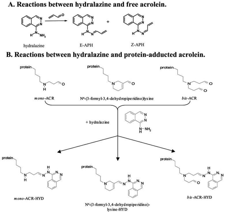
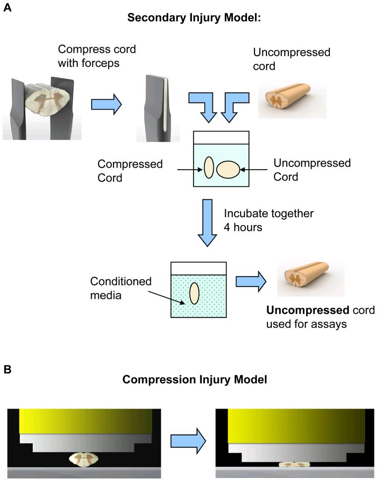
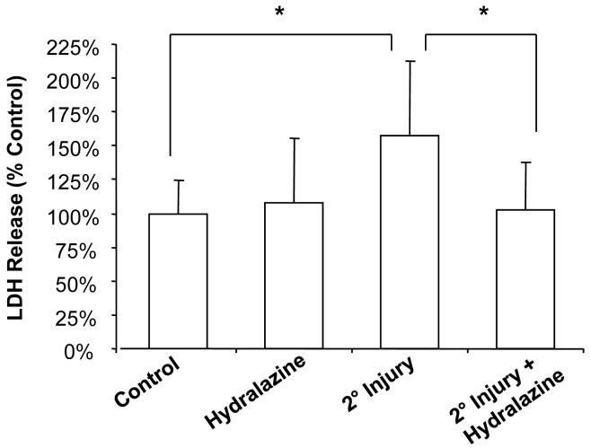
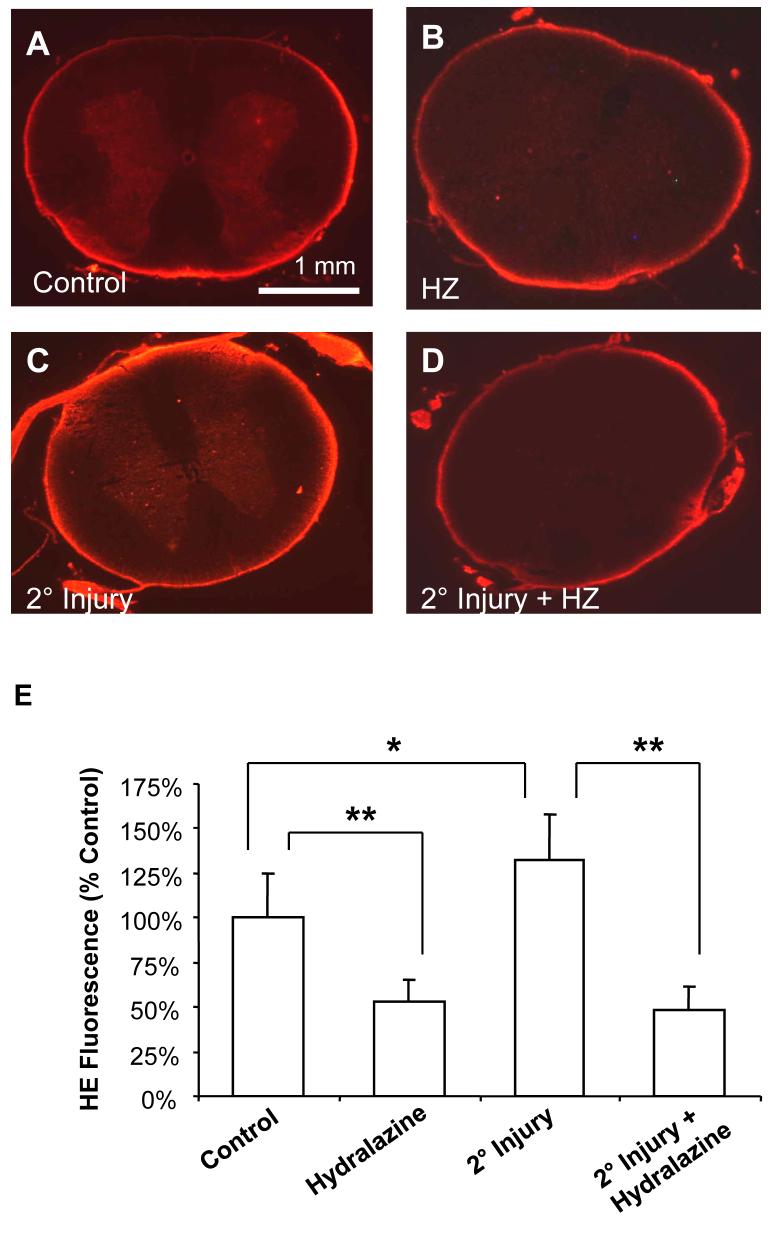
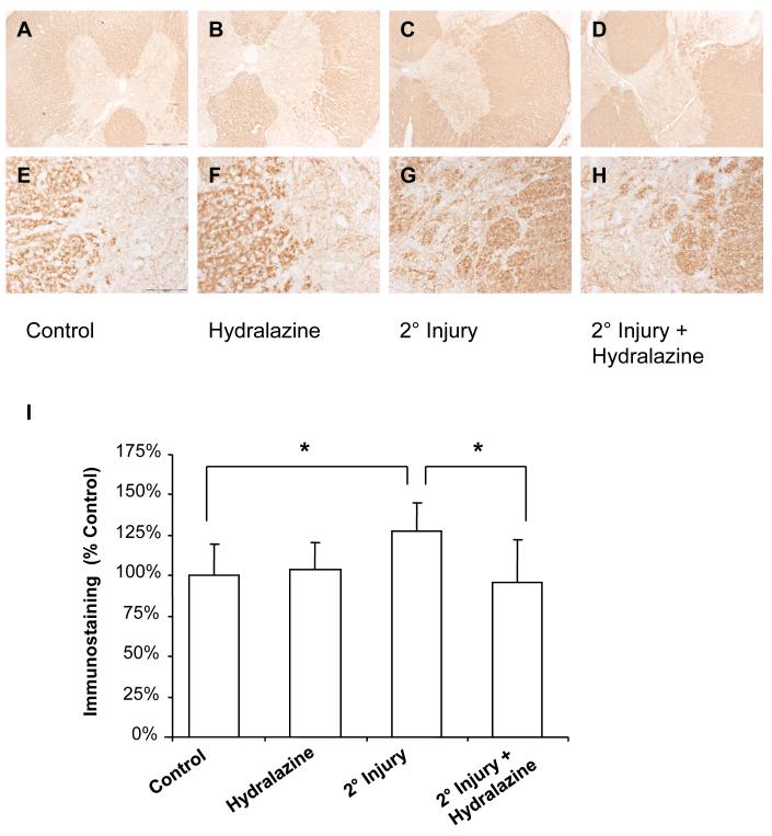
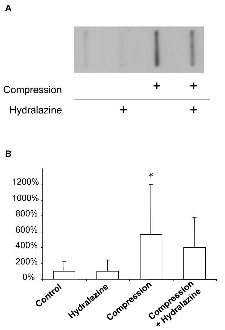
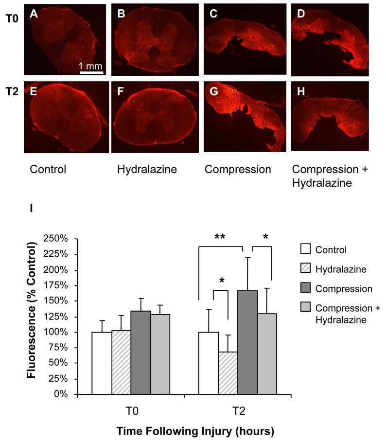
The pathophysiology of spinal cord injury (SCI) is characterized by the initial, primary injury followed by secondary injury processes in which oxidative stress is a critical component. Secondary injury processes not only exacerbate pathology at the site of primary injury, but also result in spreading of injuries to the adjacent, otherwise healthy tissue. The lipid peroxidation byproduct acrolein has been implicated as one potential mediator of secondary injury. To further and rigorously elucidate the role of acrolein in secondary injury, a unique ex vivo model is utilized to isolate the detrimental effects of mechanical injury from toxins such as acrolein that are produced endogenously following SCI. We demonstrate that (i) acrolein-Lys adducts are capable of diffusing from compressed tissue to adjacent, otherwise uninjured tissue; (ii) secondary injury by itself produces significant membrane damage and increased superoxide production; and (iii) these injuries are significantly attenuated by the acrolein scavenger hydralazine. Furthermore, hydralazine treatment results in significantly less membrane damage 2 h following compression injury, but not immediately after. These findings support our hypothesis that, following SCI, acrolein is increased to pathologic concentrations, contributes significantly to secondary injury, and thus represents a novel target for scavenging to promote improved recovery.


Figures

Figure 1
Figure 1. Chemistry of Reaction of Hydralazine and Acrolein
This illustrates the chemistry of reaction of hydralazine with free acrolein (A) and protein-adducted acrolein (B). This figure is reproduced with permission from the original manuscript (Kaminskas et al, 2004b).
Figure 2
Figure 2. Injury models
A. In the unique model of secondary injury, this illustrates the methods that were used to isolate secondary injury from mechanical injury. In this model, endogenous toxins diffuse from the compressed form, forming a “conditioned media,” in which an otherwise uninjured spinal cord segment is being bathed. B. In the compression injury model, this illustrates the compression rod that was used to induce compression injury in isolated spinal cord. The ventral side of the spinal cord was on top, and the spinal cord was compressed 70% (i.e., to 30% its original diameter) at rate of 5 mm/s.
Figure 3
Figure 3. Membrane permeability to LDH following secondary injury
Membrane permeability was assessed by release of LDH (140 kD), an intracellular enzyme that leaks out of damaged cells. Spinal cord segments were incubated for 3 hours in each of the treatment groups. The compressed cords were removed and the remaining segments rinsed and placed in fresh medium. After one additional hour, samples were taken from the medium bathing spinal cord and assayed for LDH using the TOX-7 kit. Results are expressed as % control values ± SD (n = 6). Secondary injury significantly increased permeability to LDH, an effect that was attenuated by hydralazine. One-way repeated measures ANOVA and Post Hoc Newman Keul's test were used for statistical analysis. *P<0.05.
Figure 4
Figure 4. Superoxide production following secondary injury
Superoxide production was detected by increased fluorescence following staining with HE. Representative images are shown for A) Control, B) 500 μM hydralazine (HZ), C) secondary injury, or D) secondary injury plus 500 μM hydralazine. Notice the increased fluorescence intensity following secondary injury (C). This effect is most obvious in the grey matter, and is reduced by treatment with hydralazine (B, D). Sub-pial fluorescence can probably be attributed to glial cells as well as artifactual oxidation of HE. E) Fluorescence was quantified using Image J (NIH), and is expressed as percent control values ± SD (n = 6). One-way ANOVA and Post Hoc Newman Keul's test were used for statistical analysis. *P<0.05, ** P<0.001 (compared to control).
Figure 5
Figure 5. IHC for acrolein-lys adducts
Acrolein-lys adducts were detected by immunohistochemistry. Representative images are shown (A-H). Scale bar = 500 μm for A-D, and 100 μm for E-H. Staining intensity was quantified using Image J (NIH) and is expressed as % control values ± SD (n = 4) (I). Secondary injury resulted in a slight, but statistically significant, increase in immunostaining. One-way repeated measures ANOVA and Post Hoc Newman Keul's test were used for statistical analysis. *P<0.05.
Figure 6
Figure 6. Immunoblotting for acrolein-lys adducts
Acrolein-lys adducts were detected in the Kreb's solution bathing tissue using a Bio-Dot SF Microfiltration Apparatus. A representative blot is shown (A). Relative densities were quantified using Image J (NIH) and expressed as % control values ± SD (n = 8) (B). Acrolein-lys adducts were significantly increased in Kreb's solution following compression injury, an effect that tended to be reduced by hydralazine treatment. Oneway repeated measures ANOVA and Post Hoc Newman Keul's test were used for statistical analysis. *P<0.05.
Figure 7
Figure 7. Permeability to TMR following compression injury
Membrane integrity following compression was assessed using tetramethyl rhodamine dextran (TMR, 10 kD), a hydrophilic dye that is excluded from cells with an intact membrane. Representative images are shown at 2 hours (E-H) and immediately after compression injury (A-D). Notice the increase in fluorescence intensity in compressed spinal cord compared to controls. Fluorescence was significantly reduced by treatment with hydralazine at 2 hours in both compressed and uninjured spinal cord. Immediately following injury, hydralazine had no effect on fluorescence. Fluorescence intensity was quantified using Image J (NIH) and is expressed as % control values ± SD (n = 5) (I). One-way repeated measures ANOVA and Post Hoc Newman Keul's test were used for statistical analysis. *P<0.05, ** P<0.001.

References

    1. Adams JD, Jr., Klaidman LK. Acrolein-induced oxygen radical formation. Free Radic. Biol. Med. 1993;15:187–193. - PubMed
    1. Bindokas VP, Jordan J, Lee CC, Miller RJ. Superoxide production in rat hippocampal neurons: selective imaging with hydroethidine. J. Neurosci. 1996;16:1324–1336. - PMC - PubMed
    1. Braughler JM, Hall ED. Central nervous system trauma and stroke I. Biochemical considerations for oxygen radical formation and lipid peroxidation. Free Radic. Biol. Med. 1989;6:289–301. - PubMed
    1. Burcham PC, Pyke SM. Hydralazine inhibits rapid acrolein-induced protein oligomerization: role of aldehyde scavenging and adduct trapping in cross-link blocking and cytoprotection. Mol. Pharmacol. 2006;69:1056–1065. - PubMed
    1. Burcham PC, Kerr PG, Fontaine F. The antihypertensive hydralazine is an efficient scavenger of acrolein. Redox Rep. 2000;5:47–49. - PubMed

Publication types