

Interlobular and intralobular mammary stroma: genotype may not reflect phenotype
- PMID: 18710550
- PMCID: PMC2529294
- DOI: 10.1186/1471-2121-9-46
Interlobular and intralobular mammary stroma: genotype may not reflect phenotype
Abstract
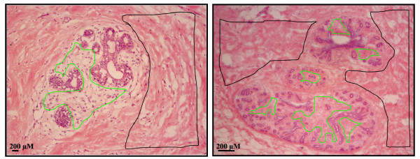
Background: The normal growth and function of mammary epithelial cells depend on interactions with the supportive stroma. Alterations in this communication can lead to the progression or expansion of malignant growth. The human mammary gland contains two distinctive types of fibroblasts within the stroma. The epithelial cells are surrounded by loosely connected intralobular fibroblasts, which are subsequently surrounded by the more compacted interlobular fibroblasts. The different proximity of these fibroblasts to the epithelial cells suggests distinctive functions for these two subtypes. In this report, we compared the gene expression profiles between the two stromal subtypes.
Methods: Fresh normal breast tissue was collected from reduction mammoplasty patients and immediately placed into embedding medium and frozen on dry ice. Tissue sections were subjected to laser capture microscopy to isolate the interlobular from the intralobular fibroblasts. RNA was prepared and subjected to microarray analysis using the Affymetrix Human Genome U133 GeneChip. Data was analyzed using the Affy and Limma packages available from Bioconductor. Findings from the microarray analysis were validated by RT-PCR and immunohistochemistry.
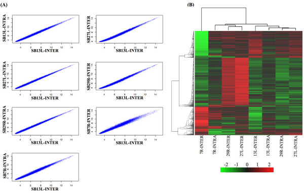
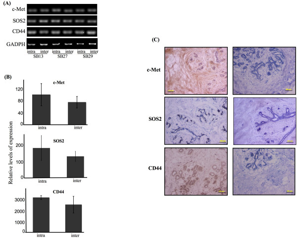
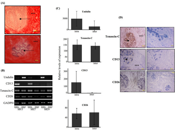
Results: No statistically significant difference was detected between the gene expression profiles of the interlobular and intralobular fibroblasts by microarray analysis and RT-PCR. However, for some of the genes tested, the protein expression patterns between the two subtypes of fibroblasts were significantly different.
Conclusion: This study is the first to report the gene expression profiles of the two distinct fibroblast populations within the human mammary gland. While there was no significant difference in the gene expression profiles between the groups, there was an obvious difference in the expression pattern of several proteins tested. This report also highlights the importance of studying gene regulation at both the transcriptional and post-translational level.
Figures
References
-
- Jemal A, Siegel R, Ward E, Murray T, Xu J, Thun MJ. Cancer statistics, 2007. CA Cancer J Clin. 2007;57:43–66. - PubMed
-
- Patanaphan V, Salazar OM, Risco R. Breast cancer: metastatic patterns and their prognosis. South Med J. 1988;81:1109–1112. - PubMed
-
- Gache C, Berthois Y, Cvitkovic E, Martin PM, Saez S. Differential regulation of normal and tumoral breast epithelial cell growth by fibroblasts and 1,25-dihydroxyvitamin D3. Breast Cancer Res Treat. 1999;55:29–39. - PubMed
-
- van Roozendaal KE, Klijn JG, van Ooijen B, Claassen C, Eggermont AM, Henzen-Logmans SC, Foekens JA. Differential regulation of breast tumor cell proliferation by stromal fibroblasts of various breast tissue sources. Int J Cancer. 1996;65:120–125. - PubMed
Publication types
MeSH terms
Substances
Grants and funding
LinkOut - more resources
Full Text Sources
Molecular Biology Databases
