Case Reports
. 2008 Nov 25:1242:252-62.
doi: 10.1016/j.brainres.2008.07.088. Epub 2008 Jul 30.

Neural and behavioral correlates of drawing in an early blind painter: a case study

Affiliations
Case Reports

Neural and behavioral correlates of drawing in an early blind painter: a case study

Amir Amedi et al. Brain Res. .

Abstract

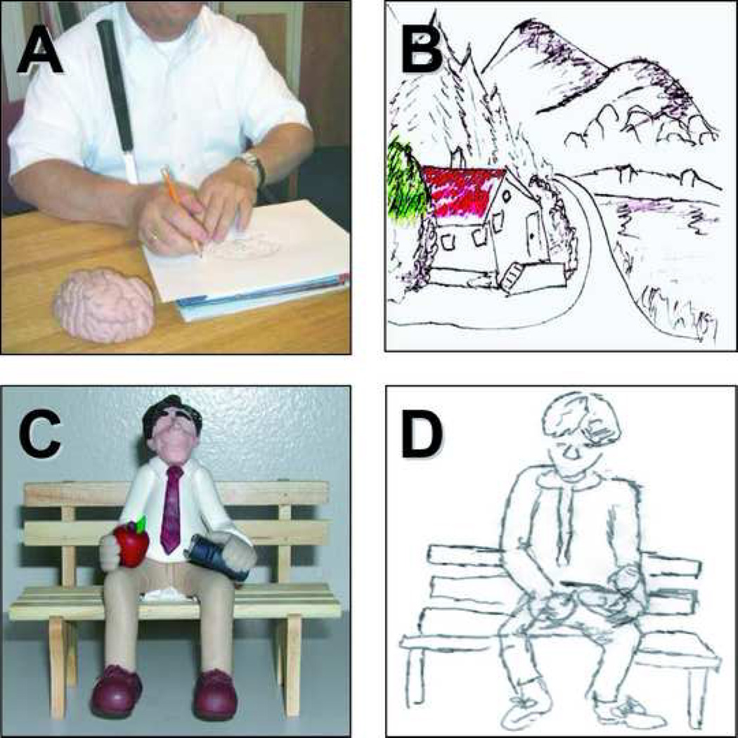
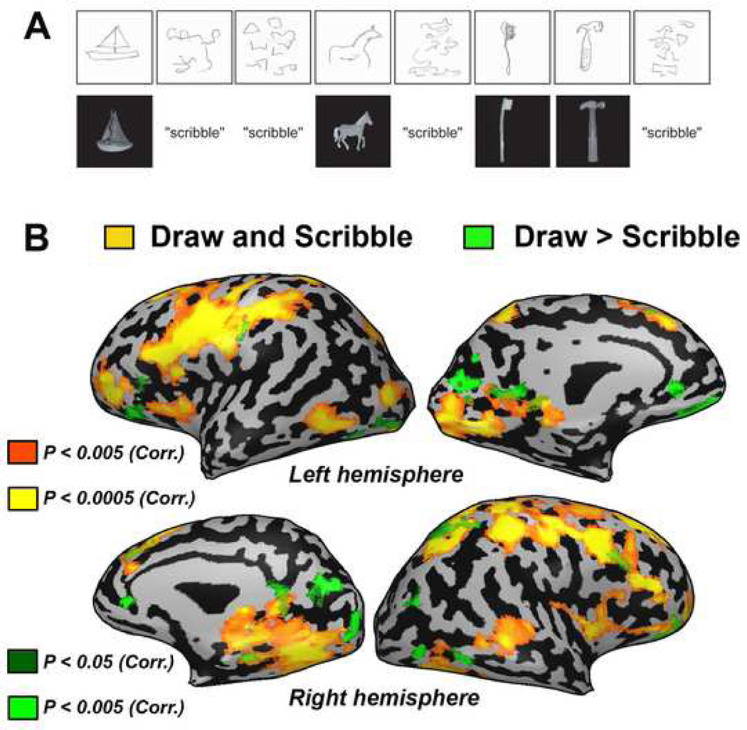
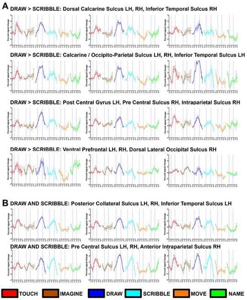
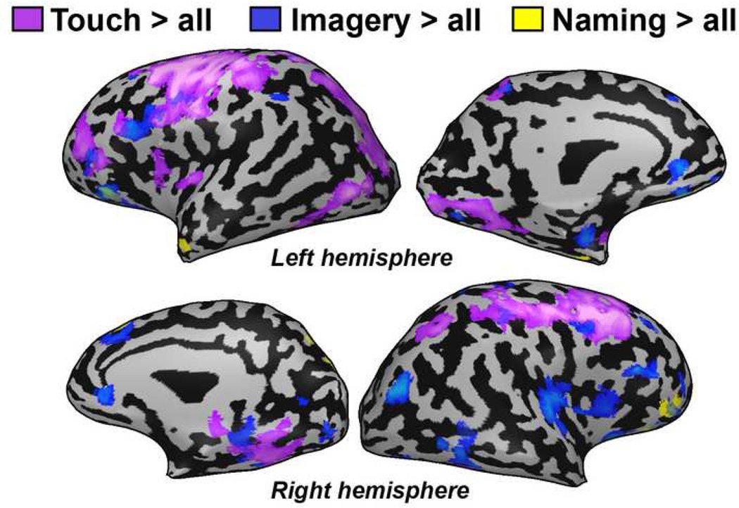
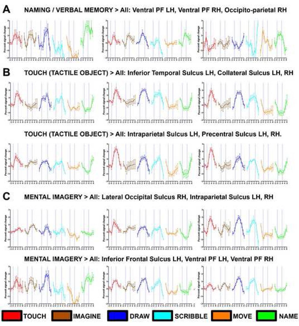
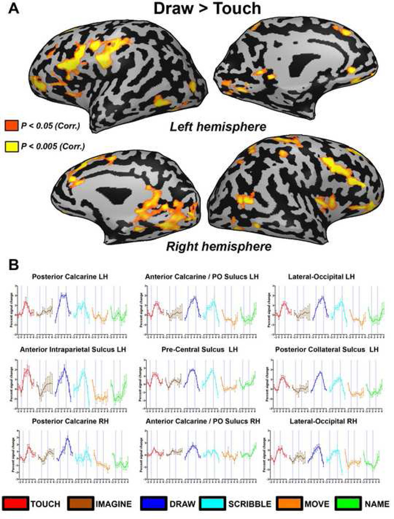
Humans rely heavily on vision to identify objects in the world and can create mental representations of the objects they encounter. Objects can also be identified and mentally represented through haptic exploration. However, it is unclear whether prior visual experience is necessary to generate these internal representations. Subject EA, an early blind artist, provides insight into this question. Like other blind individuals, EA captures the external world by touch. However, he is also able to reveal his internal representations through highly detailed drawings that are unequivocally understandable by a sighted person. We employed fMRI to investigate the neural correlates associated with EA's ability to transform tactilely explored three-dimensional objects into drawings and contrasted these findings with a series of control conditions (e.g. nonsensical scribbling as a sensory-motor control). Activation during drawing (compared to scribbling) occurred in brain areas normally associated with vision, including the striate cortex along with frontal and parietal cortical regions. Some of these areas showed overlap when EA was asked to mentally imagine the pictures he had to draw (albeit to a lesser anatomical extent and signal magnitude). These results have important implications as regards our understanding of the ways in which tactile information can generate mental representations of shapes and scenes in the absence of normal visual development. Furthermore, these findings suggest the occipital cortex plays a key role in supporting mental representations even without prior visual experience.


Figures

Figure 1
Figure 1. Examples of subject EA’s drawing abilities
A) EA drawing a novel object (a model of the brain) using a pencil and paper and a specially designed rubberized writing tablet (Sewell raised line drawing kit). This technique allows him to generate relief images that he can subsequently detect and explore tactilely. (B) The themes of his drawings and paintings vary and include both tactile and non-tactile subjects. The drawing shows a landscape scene and illustrates how he applies colors to his paintings. His paintings often contain vibrant color, and he uses shading, depth cues, and perspective akin to that employed by sighted artists. (C) Example of a complex and novel object which EA had never encountered. Once EA explored the object by touch for a few minutes, he was able to render a very accurate drawing of the object (D).
Figure 2
Figure 2. Neuroimaging data of subject EA for drawing and scribbling objects
A) Example of behavioral data collected from a scanning run. Subject EA’s sketches (above) are shown compared to the object tactilely explored or in response to the control scribble condition (below). (B) Drawing versus scribbling contrast presented on a full inflated cortical reconstruction of EA's brain. Activation was found in a network of posterior occipital (including the calcarine sulcus), occipito-temporal, occipito-parietal and prefrontal areas.
Figure 3
Figure 3. Time courses from selected regions of interest (ROIs) for Drawing vs. Scribbling (A) and Drawing and Scribbling vs. Baseline (B)
For demonstrative purposes, we also present the time-course of all experimental conditions in different ROIs using the peak voxel in the smoothed volume of each ROI. (A) Several ROIs in the occipital cortex show robust and selective activation for drawing objects with a typical hemodynamic response. Each ROI (corresponding to the green scale clusters shown in Figure 2) can be identified by its header. A similar pattern was also found in clusters within the parietal cortex. Robust activation for drawing was also found in prefrontal cortex, but in these areas there was also relatively robust activation for imagery and naming/verbal memory (in spite of the fact that these conditions were ignored in the contrast used to select the ROIs in this test). (B) ROIs of peak activation for clusters significantly activated by drawing and scribbling versus baseline (similar to those shown in Figure 2 in red to yellow color scale). The time courses are depicted by the following color index: tactile objects (red), mental imagery (brown), drawing (blue), scribbling (cyan), motor control (orange) and naming/verbal memory (green).
Figure 4
Figure 4. Neuroimaging data of subject EA for object recognition by touch (purple), mental imagery (blue) and naming (yellow)
Each control condition was contrasted with all the other control conditions in order to exclude non-specific effect (e.g. the purple, touch cluster are defined by the contrast: touch > imagery, naming and sensory-motor control). Activation as a result of the recognition of different features of the object by touch and mental imagery of the object drawn included many similar areas in prefrontal, parietal and occipital areas.
Figure 5
Figure 5. Time courses from selected ROIs for naming (A) object recognition by touch (B) and mental imagery (C)
A) Several ROIs in the prefrontal and parietal cortex show robust activation for recalling the names of objects from memory. Activation was not very selective as these clusters also showed activation for mental imagery, drawing and tactile objects. (B) Time courses for tactile object exploration. All ROIs showed robust activation for both the drawing and scribbling tasks. (C) Time courses for mental imagery of the explored objects (which EA had to draw next). Most of these ROIs also showed activation for drawing and touching objects.
Figure 6
Figure 6
Direct comparison of drawing versus tactilely exploring objects. (A) The contrast of drawing versus touching objects is presented on a full inflated cortical reconstruction of EA's brain. Activation was found mainly in several posterior ventral occipital areas (including the calcarine sulcus) and prefrontal cortex. Some activation was also found in parietal cortex and the Insula. (B) Time courses from selected regions of interest (ROIs) for drawing versus touching objects that were highlighted in (A).

References

    1. Amedi A, Raz N, Pianka P, Malach R, Zohary E. Early 'visual' cortex activation correlates with superior verbal memory performance in the blind. Nat Neurosci. 2003;6:758–766. - PubMed
    1. Amedi A, von Kriegstein K, van Atteveldt NM, Beauchamp MS, Naumer MJ. Functional imaging of human crossmodal identification and object recognition. Exp Brain Res. 2005;166:559–571. - PubMed
    1. Annoni JM, Devuyst G, Carota A, Bruggimann L, Bogousslavsky J. Changes in artistic style after minor posterior stroke. J Neurol Neurosurg Psychiatry. 2005;76:797–803. - PMC - PubMed
    1. Binkofski F, Buccino G, Posse S, Seitz RJ, Rizzolatti G, Freund H. A fronto-parietal circuit for object manipulation in man: evidence from an fMRI-study. Eur J Neurosci. 1999;11:3276–3286. - PubMed
    1. Boynton GM, Engel SA, Glover GH, Heeger DJ. Linear systems analysis of functional magnetic resonance imaging in human V1. J Neurosci. 1996;16:4207–4221. - PMC - PubMed

Publication types