. 2008 Sep 1;205(9):1975-81.
doi: 10.1084/jem.20080308. Epub 2008 Aug 18.

CD4+ FoxP3+ regulatory T cells confer infectious tolerance in a TGF-beta-dependent manner

Affiliations

CD4+ FoxP3+ regulatory T cells confer infectious tolerance in a TGF-beta-dependent manner

John Andersson et al. J Exp Med. .

Abstract

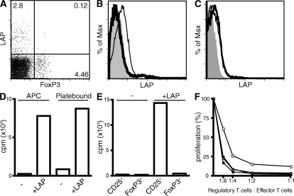
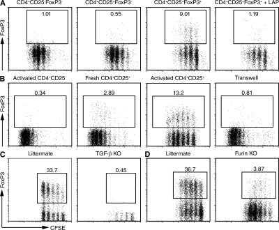
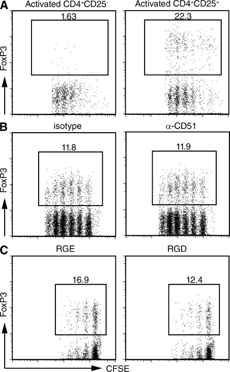
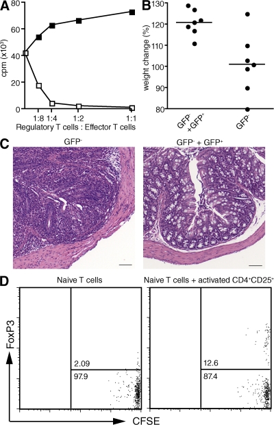
CD4(+)FoxP3(+) regulatory T (T reg) cells comprise a separate lineage of T cells that are essential for maintaining immunological tolerance to self. The molecular mechanism(s) by which T reg cells mediate their suppressive effects remains poorly understood. One molecule that has been extensively studied in T reg cell suppression is transforming growth factor (TGF)-beta, but its importance remains controversial. We found that TGF-beta complexed to latency-associated peptide (LAP) is expressed on the cell surface of activated but not resting T reg cells. T reg cell LAP-TGF-beta plays an important role in the suppression of the proliferation of activated T cells, but it is not required for the suppression of naive T cell activation. More importantly, T reg cell-derived TGF-beta could generate de novo CD4(+)FoxP3(+) T cells in vitro from naive precursors in a cell contact-dependent, antigen-presenting cell-independent and alpha(V) integrin-independent manner. The newly induced CD4(+)FoxP3(+) T cells are suppressive both in vitro and in vivo. Transfer of activated antigen-specific T reg cells with naive antigen-specific responder T cells to normal recipients, followed by immunization, also results in induction of FoxP3 expression in the responder cells. T reg cell-mediated generation of functional CD4(+)FoxP3(+) cells via this TGF-beta-dependent pathway may represent a major mechanism as to how T reg cells maintain tolerance and expand their suppressive abilities.


Figures

Figure 1.
Figure 1.
LAP is expressed by activated T reg cells, and TGF-β is required to suppress CD4+CD25+FoxP3 T cells. (A) Expression of LAP and FoxP3 on freshly isolated lymph node cells from C57BL/6 mice. (B) CD4+CD25 (thick line histogram) and CD4+CD25+ T cells (thin line histogram) were activated for 3 d with plate-bound anti-CD3 and IL-2 and analyzed for LAP expression. Isotype control is depicted with a solid gray histogram. (C) Activated CD4+CD25+ T cells were incubated with (thick line histogram) or without (thin line histogram) 20 μg/ml LAP for 2 h at 4°C and analyzed for LAP expression. Isotype control is depicted with a solid gray histogram. (D) CD4+CD25+ T cells (5 × 10⁴/well) from lymph nodes of C57BL/6 mice were activated with plate-bound anti-CD3 or with irradiated T cell–depleted splenocytes (5 × 10⁴/well) and 0.25 μg/ml of soluble anti-CD3 in the presence or absence of LAP. [3H]TdR incorporation was determined after 72 h of culture. (E) CD4+CD25+ from C57BL/6 mice or CD4+CD25+ GFP+ T cells from C57BL/6 FoxP3-GFP knock-in mice were cultured together with irradiated T cell–depleted splenocytes (5 × 10⁴/well) and 0.25 μg/ml of soluble anti-CD3 in the presence or absence of LAP. [3H]TdR incorporation was determined after 72 h of culture. (F) CD4+CD25 C57BL/6 T cells (5 × 10⁴/well) were co-cultured with CD4+CD25+ T cells from C57BL/6 mice (squares) or with CD4+CD25+GFP+ T cells from C57BL/6 FoxP3-GFP knock-in mice (circles) in the presence of irradiated T cell–depleted splenocytes (5 × 10⁴/well) and 0.25 μg/ml of soluble anti-CD3 with (open symbols) or without (closed symbols) 12.5 μg/ml LAP. [3H]TdR incorporation was determined after 72 h of culture. One representative experiment of at least three is shown (A–F).
Figure 2.
Figure 2.
T reg cells confer infectious tolerance in a TGF-β–dependent manner. (A) Activated CD4+CD25FoxP3, CD4+CD25+FoxP3, and CD4+CD25+FoxP3+ T cells were co-cultured with CFSE-labeled 5CC7 TCR transgenic RAG−/− T cells, 2 μg/ml anti-CD3, splenic DCs, and IL-2 (100 U/ml) in the absence or presence of LAP. (B) Activated CD4+CD25+ and freshly isolated CD4+CD25+ were co-cultured with CFSE-labeled 5CC7 TCR transgenic RAG−/− T cells, 2 μg/ml anti-CD3, splenic DCs, and IL-2 (100 U/ml) (left panels). Activated CD4+CD25+ T cells were co-cultured with the responders or separated from the responders in a transwell (right panel). (C) Activated CD4+CD25+ T cells from the thymus of 2-wk-old TGF-β1−/− mice or from littermate controls were co-cultured with CFSE-labeled 5CC7 TCR transgenic RAG−/− T cells. (D) Activated CD4+FoxP3+ T cells from peripheral lymph nodes of CD4cre-furf/f mice or littermate controls were co-cultured with CFSE-labeled 5CC7 TCR transgenic RAG−/− T cells. Flow cytometric analysis was used to determine the level of FoxP3 expression after 4 d of the co-culture. All dot plots were gated on CFSE+ cells as shown in Fig. S2. One representative experiment out of at least two experiments is shown (A–D). Fig. S2 is available at http://www.jem.org/cgi/content/full/jem.20080308/DC1.
Figure 3.
Figure 3.
The induction of FoxP3 expression by activated T reg cells is APC and αv integrin independent. (A) CD4+CD25 (left) and CD4+CD25+ (right) T cells from B10.A mice were activated for 4 d with plate-bound anti-CD3 and IL-2 (100 U/ml). The activated T cells were then co-cultured for an additional 4 d with an equal number of CFSE-labeled CD4+ T cells from 5CC7 TCR transgenic RAG−/− mice in the presence of plate-bound anti-CD3 and IL-2. (B and C) CD4+CD25+ T cells were activated with plate-bound anti-CD3 and IL-2 for 4 d in the absence (left) or the presence (right) of blocking anti-αv monoclonal antibodies (50 μg/ml), RGE peptide, or RGD peptide (10 μg/ml). The activated cells were then co-cultured with CFSE-labeled 5CC7 TCR transgenic T cells co-cultured in the presence or absence of the same inhibitors used in the first culture. FoxP3 expression was determined on day 4 of the co-culture. One representative experiment out of at least three is shown (A–C).
Figure 4.
Figure 4.
Induced FoxP3+ T cells are suppressive in vitro and in vivo. (A) T reg cell–induced CD4+FoxP3+ T cells (open squares) were isolated as in Fig. S5 and tested for suppressive ability by co-culturing them with CD4+CD25 T cells (5 × 10⁴/well) in the presence of irradiated T cell–depleted splenocytes (5 × 10⁴/well) and 0.25 μg/ml of soluble anti-CD3. Activated CD4+CD25GFP T cells (closed squares) from C57BL/6 FoxP3-GFP mice were used as controls. Proliferation was measured by the incorporation of [3H]TdR after 72 h of culture. (B) CD4+GFP T cells (5 × 10⁵) from C57BL/6 FoxP3-GFP knock-in mice were transferred into C57BL/6 RAG−/− mice with or without 1.5 × 10⁵ T reg cell–induced FoxP3+ T cells isolated as described in Fig. S5. The weight of all recipients was measured at 6 wk after cell transfer. P < 0.006 using Student's t test. (C) Hematoxylin and eosin–stained tissue sections from the colon of C57BL/6 RAG−/− mice that received CD4+FoxP3 T cells with (right) or without (left) the addition of T reg cell–induced CD4+FoxP3+ T cells. Bar, 100 μm. (D) CFSE-labeled CD4+CD25CD44 HA TCR transgenic T cells (0.5 × 10⁶) were transferred into B10.D2 mice with or without activated HA-specific TCR transgenic T reg cells (2.5 × 10⁶). 1 d later, all recipients were immunized with HA peptide in IFA, and draining lymph node cells were analyzed for FoxP3 expression 48 h after transfer. One representative experiment out of at least two is shown (A–D). Fig. S5 is available at http://www.jem.org/cgi/content/full/jem.20080308/DC1.

References

    1. Khattri, R., T. Cox, S.A. Yasayko, and F. Ramsdell. 2003. An essential role for Scurfin in CD4+CD25+ T regulatory cells. Nat. Immunol. 4:337–342. - PubMed
    1. Hori, S., T. Nomura, and S. Sakaguchi. 2003. Control of regulatory T cell development by the transcription factor Foxp3. Science. 299:1057–1061. - PubMed
    1. Fontenot, J.D., M.A. Gavin, and A.Y. Rudensky. 2003. Foxp3 programs the development and function of CD4+CD25+ regulatory T cells. Nat. Immunol. 4:330–336. - PubMed
    1. Brunkow, M.E., E.W. Jeffery, K.A. Hjerrild, B. Paeper, L.B. Clark, S.A. Yasayko, J.E. Wilkinson, D. Galas, S.F. Ziegler, and F. Ramsdell. 2001. Disruption of a new forkhead/winged-helix protein, scurfin, results in the fatal lymphoproliferative disorder of the scurfy mouse. Nat. Genet. 27:68–73. - PubMed
    1. Bennett, C.L., J. Christie, F. Ramsdell, M.E. Brunkow, P.J. Ferguson, L. Whitesell, T.E. Kelly, F.T. Saulsbury, P.F. Chance, and H.D. Ochs. 2001. The immune dysregulation, polyendocrinopathy, enteropathy, X-linked syndrome (IPEX) is caused by mutations of FOXP3. Nat. Genet. 27:20–21. - PubMed

Publication types

Substances