. 2008 Oct;28(20):6149-59.
doi: 10.1128/MCB.00220-08. Epub 2008 Aug 18.

Regulation of the endosomal SNARE protein syntaxin 7 by colony-stimulating factor 1 in macrophages

Affiliations

Regulation of the endosomal SNARE protein syntaxin 7 by colony-stimulating factor 1 in macrophages

Adrian Achuthan et al. Mol Cell Biol. 2008 Oct.

Abstract

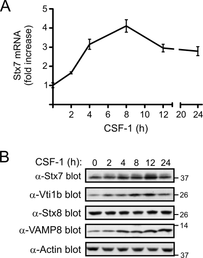
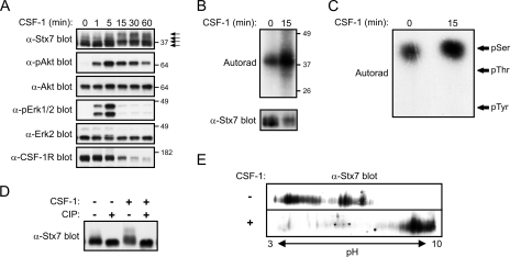
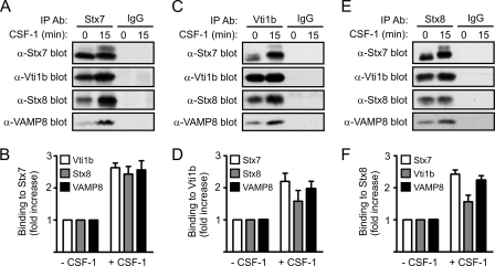
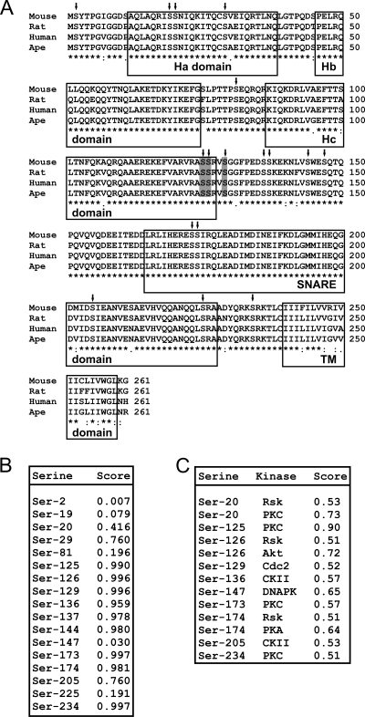
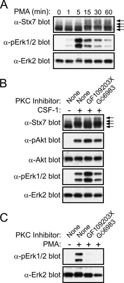
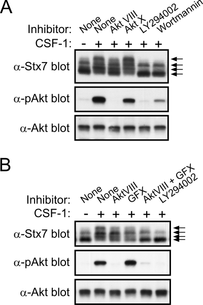
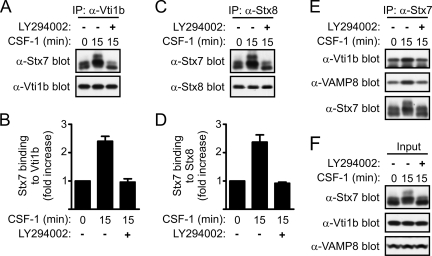
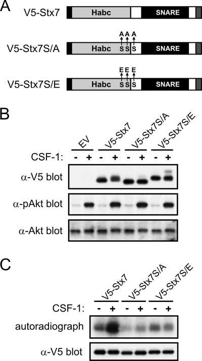
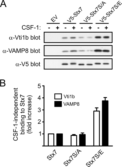
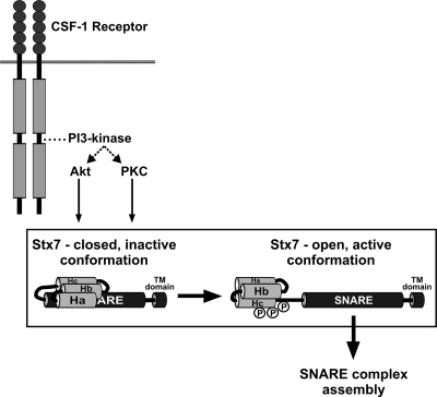
Colony-stimulating factor 1 (CSF-1) is the main growth factor controlling the development of macrophages from myeloid progenitor cells. However, CSF-1 also regulates some of the key effector functions of macrophages (e.g., phagocytosis and cytokine secretion). The endosomal SNARE protein syntaxin 7 (Stx7) regulates vesicle trafficking events involved in phagocytosis and cytokine secretion. Therefore, we investigated the ability of CSF-1 to regulate Stx7. CSF-1 upregulated Stx7 expression in primary mouse macrophages; it also upregulated expression of its SNARE partners Vti1b and VAMP8 but not Stx8. Additionally, CSF-1 induced the rapid serine phosphorylation of Stx7 and enhanced its binding to Vti1b, Stx8, and VAMP8. Bioinformatics analysis and results from experiments with kinase inhibitors suggested the CSF-1-induced phosphorylation of Stx7 was mediated by protein kinase C and Akt in response to phosphatidylinositol 3-kinase activation. Based on mutagenesis studies, CSF-1 appeared to increase the binding of Stx7 to its SNARE partners by inducing the phosphorylation of serine residues in the Habc domain and/or "linker" region of Stx7. Thus, CSF-1 is a key regulator of Stx7 expression and function in macrophages. Furthermore, the effects of CSF-1 on Stx7 may provide a mechanism for the regulation of macrophage effector functions by CSF-1.


Figures

FIG. 1.
FIG. 1.
Regulation of Stx7 expression by CSF-1. (A) Mouse bone marrow-derived macrophages were deprived of CSF-1 for 16 h before being stimulated with CSF-1 for the time indicated. Total RNA was then extracted and reverse transcribed into cDNA, which was then subjected to quantitative real-time PCR. 18S rRNA was used as the internal control. Stx7 mRNA levels are relative to its expression in the absence of CSF-1, which was given an arbitrary value of 1.0. Stx7 mRNA levels (mean ± standard error) at each time point were measured in triplicate, and the data are representative of three experiments. (B) Macrophages were cultured as for panel A and cell lysates subsequently subjected to Western blotting with the indicated antibodies. The membrane was also probed with an antiactin antibody to assess loading. The positions of molecular mass markers (in kDa) are indicated on the right. α-Stx7, anti-Stx7; other antibodies are similarly indicated.
FIG. 2.
FIG. 2.
CSF-1-induced phosphorylation of Stx7 in macrophages. (A) Mouse bone marrow-derived macrophages were deprived of CSF-1 for 16 h before being stimulated with CSF-1 for the time indicated. The cells were then lysed and the lysates subsequently subjected to Western blotting with the indicated antibodies. The positions of molecular mass markers (in kDa) and the different Stx7 isoforms (arrows) are indicated on the right. α-Stx7, anti-Stx7; other antibodies are similarly indicated. (B and C) Macrophages that had been metabolically labeled with [32P]orthophosphate were stimulated with CSF-1 for 15 min and then lysed. (B) Stx7 was immunoprecipitated from the lysates using anti-Stx7 antibodies and the immunoprecipitates subsequently subjected to autoradiography (upper panel) or Western blotting with an anti-Stx7 antibody (lower panel). (C) Stx7 that had been immunoprecipitated from the [32P]orthophosphate-labeled macrophages in panel B was excised from the PVDF membrane and subjected to phosphoamino acid analysis. The positions of phosphoamino acid standards (pSer, phosphoserine; pThr, phosphothreonine; pTyr, phosphotyrosine) are indicated on the right. (D) Macrophages were deprived of CSF-1 for 16 h before being stimulated with CSF-1 for 15 min and then lysed in the absence of phosphatase inhibitors. Aliquots of the lysates were incubated with CIP or in reaction buffer alone for 30 min at 37°C. The lysates were then subjected to Western blotting with an anti-Stx7 antibody. (E) Lysates of macrophages that had been left unstimulated or which had been stimulated with CSF-1 for 15 min were subjected to two-dimensional SDS-PAGE analysis followed by Western blotting with an anti-Stx7 antibody.
FIG. 3.
FIG. 3.
Effects of CSF-1 on Stx7 SNARE complexes in macrophages. Mouse bone marrow-derived macrophages were deprived of CSF-1 for 16 h before being stimulated with CSF-1 for 15 min. Following cell lysis, Stx7 (A and B), Vti1b (C and D), and Stx8 (E and F) were immunoprecipitated from the lysates using anti-Stx7, anti-Vti1b, and anti-Stx8 antibodies (α-Stx7, α-Vti1b, and α-Stx8), respectively. Lysates were also immunoprecipitated with irrelevant control antibodies. (A, C, and E). The immunoprecipitates were subsequently subjected to Western blotting with the indicated antibodies. (B, D, and F) Quantified data are presented as the increase in SNARE protein binding following CSF-1 stimulation relative to that in unstimulated macrophages. Data represent the means (± standard errors) of at least three experiments.
FIG. 4.
FIG. 4.
Predicted serine phosphorylation sites in Stx7. (A) The amino acid sequences of mouse, rat, human, and ape Stx7 were aligned using the ClustalW algorithm. Serine residues that are conserved across all four species are indicated with inverted arrows. Ser-125, Ser-126, and Ser-129 are shaded in gray. The Ha, Hb, Hc, SNARE, and transmembrane (TM) domains are boxed. (B) The amino acid sequence of mouse Stx7 was analyzed using the NetPhos 2.0 software program. Predicted serine phosphorylation sites in Stx7 and their corresponding probability scores are shown. (C) The amino acid sequence of mouse Stx7 was analyzed using the NetPhosK 1.0 software program. Predicted serine phosphorylation sites in Stx7, the protein kinase predicted to phosphorylate the site, and their corresponding probability scores (>0.50) are shown.
FIG. 5.
FIG. 5.
Phosphorylation of Stx7 by PKC in macrophages. (A) Mouse bone marrow-derived macrophages were deprived of CSF-1 for 16 h before being stimulated with 100 nM PMA for the time indicated. The cells were then lysed and the lysates subsequently subjected to Western blotting with the indicated antibodies. The different Stx7 isoforms are indicated by arrows on the right. α-Stx7, -pErk1/2, and -Erk2 represent anti-Stx7, -pErk1/2, and -Erk2. (B) Macrophages were deprived of CSF-1 for 16 h before being treated with 0.1% dimethyl sulfoxide, 5 μM GF109203X, or 1 μM Gö6983 for 30 min. The macrophages were stimulated with CSF-1 for 15 min and then lysed. The lysates were subsequently subjected to Western blotting with the indicated antibodies (shown as in panel A). (C) Macrophages were deprived of CSF-1 for 16 h before being treated with 0.1% dimethyl sulfoxide, 5 μM GF109203X, or 1 μM Gö6983 for 30 min. The macrophages were stimulated with PMA for 15 min and then lysed. The lysates were subsequently subjected to Western blotting with the indicated antibodies.
FIG. 6.
FIG. 6.
Effects of Akt and PI 3-kinase inhibitors on CSF-1-induced phosphorylation of Stx7 in macrophages. (A) Mouse bone marrow-derived macrophages were deprived of CSF-1 for 16 h before being treated with 0.1% dimethyl sulfoxide, 10 μM Akt-VIII, 5 μM Akt-X, 10 μM LY294002, or 100 nM wortmannin for 30 min. The macrophages were stimulated with CSF-1 for 15 min and then lysed. The lysates were subsequently subjected to Western blotting with the indicated antibodies (anti-Stx7, anti-pAkt, and anti-Akt). The different Stx7 isoforms are indicated by arrows on the right. (B) Macrophages were deprived of CSF-1 for 16 h before being treated with 0.1% dimethyl sulfoxide, 10 μM Akt VIII, 5 μM GF109203X (GFX), 10 μM Akt VIII and 5 μM GF109203X together, or 10 μM LY294002 for 30 min. The macrophages were stimulated with CSF-1 for 15 min and then lysed. The lysates were subsequently subjected to Western blotting with the indicated antibodies.
FIG. 7.
FIG. 7.
Effects of PI 3-kinase inhibition on Stx7 SNARE complexes in macrophages. Mouse bone marrow-derived macrophages were deprived of CSF-1 for 16 h before being treated with 0.1% dimethyl sulfoxide or 10 μM LY294002 for 30 min. The macrophages were stimulated with CSF-1 for 15 min and then lysed. Vti1b (A and B), Stx8 (C and D), and Stx7 (E) were subsequently immunoprecipitated (IP) from the cell lysates with anti-Vti1b (α-Vti1b), anti-Stx8 (α-Stx8), and anti-Stx7 (α-Stx7) antibodies, respectively, and then Western blotted with the indicated antibodies. (B and D) Quantified data are presented as the increase in SNARE protein binding following CSF-1 stimulation relative to that in unstimulated macrophages. Data represent the means (± standard errors) of three experiments. (F) Western blotting of cell lysates with the indicated antibodies.
FIG. 8.
FIG. 8.
Effects of mutating Ser-125, Ser-126, and Ser-129 on the CSF-1-induced phosphorylation of Stx7. (A) Schematic representation of V5-tagged wild-type Stx7 (V5-Stx7) and V5-Stx7 in which Ser-125, Ser-126, and Ser-129 have been replaced with alanine (V5-Stx7S/A) or glutamic acid (V5-Stx7S/E). The C-terminal transmembrane anchor domain is represented by a gray filled box. (B and C) Mouse bone marrow cells were transduced with retroviruses expressing V5-Stx7, V5-Stx7S/A, or V5-Stx7S/E or were transduced with a virus containing the empty vector (EV). The cells were cultured in the presence of CSF-1 until they differentiated into adherent macrophages. (B) The macrophages were then deprived of CSF-1 for 16 h before being stimulated with CSF-1 for 15 min. The cells were lysed and the lysates subsequently subjected to Western blotting with the indicated antibodies (anti-V5, anti-pAkt, and anti-Akt). (C) Retrovirally transduced macrophages were metabolically labeled with [32P]orthophosphate before being stimulated with CSF-1 for 15 min. The cells were then lysed and the V5-tagged Stx7 proteins immunoprecipitated using anti-V5 antibodies. The immunoprecipitates were subsequently subjected to autoradiography (upper panel) and Western blotting with an anti-V5 antibody (lower panel).
FIG. 9.
FIG. 9.
Effects of mutating Ser-125, Ser-126, and Ser-129 in Stx7 on the assembly of Stx7 SNARE complexes. Mouse bone marrow cells were transduced with retroviruses expressing V5-Stx7, V5-Stx7S/A, or V5-Stx7S/E or were transduced with a virus containing the empty vector (EV). The cells were then cultured in the presence of CSF-1 until they differentiated into adherent macrophages. The macrophages were deprived of CSF-1 for 16 h before being stimulated with CSF-1 for 15 min. (A) The cells were then lysed and the V5-tagged Stx7 proteins immunoprecipitated using anti-V5 (α-V5) antibodies. The immunoprecipitates were subsequently subjected to Western blotting with the indicated antibodies (anti-Vti1b [α-Vti1b], anti-VAMP8 [α-VAMP8], and anti-V5). (B) Quantified data are presented as the increase in the CSF-1-independent binding of Vti1b and VAMP8 to V5-Stx7S/A and V5-Stx7S/E relative to their binding to V5-Stx7. Data represent the means (± standard errors) of three independent experiments.
FIG. 10.
FIG. 10.
A model for the regulation of the binding of Stx7 to its partner SNARE proteins by CSF-1. In the absence of CSF-1, Stx7 adopts predominantly a “closed” conformation by virtue of the Habc domain intramolecularly interacting with the SNARE domain, thereby suppressing the binding of Stx7 to its partner SNARE proteins. Upon activation of the CSF-1 receptor, Akt and PKC become activated, via PI 3-kinase, and phosphorylate Ser-125, Ser-126, and/or Ser-129 in the Hc domain and/or “linker” region of Stx7. This induces a conformational change in Stx7 that results in Stx7 adopting an “open” conformation with an enhanced capacity to bind to its SNARE partners (e.g., Vti1b, Stx8, and VAMP8).

Similar articles

Cited by

References

    1. Aderem, A., and D. M. Underhill. 1999. Mechanisms of phagocytosis in macrophages. Annu. Rev. Immunol. 17593-623. - PubMed
    1. Antonin, W., I. Dulubova, D. Arac, S. Pabst, J. Plitzner, J. Rizo, and R. Jahn. 2002. The N-terminal domains of syntaxin 7 and vti1b form three-helix bundles that differ in their ability to regulate SNARE complex assembly. J. Biol. Chem. 27736449-36456. - PubMed
    1. Antonin, W., C. Holroyd, D. Fasshauer, S. Pabst, G. F. Von Mollard, and R. Jahn. 2000. A SNARE complex mediating fusion of late endosomes defines conserved properties of SNARE structure and function. EMBO J. 196453-6464. - PMC - PubMed
    1. Araki, N., M. T. Johnson, and J. A. Swanson. 1996. A role for phosphoinositide 3-kinase in the completion of macropinocytosis and phagocytosis by macrophages. J. Cell Biol. 1351249-1260. - PMC - PubMed
    1. Arcaro, A., and M. P. Wymann. 1993. Wortmannin is a potent phosphatidylinositol 3-kinase inhibitor: the role of phosphatidylinositol 3,4,5-trisphosphate in neutrophil responses. Biochem. J. 296297-301. - PMC - PubMed

Publication types

MeSH terms