

Lactate stimulates vasculogenic stem cells via the thioredoxin system and engages an autocrine activation loop involving hypoxia-inducible factor 1
- PMID: 18710947
- PMCID: PMC2577432
- DOI: 10.1128/MCB.00795-08
Lactate stimulates vasculogenic stem cells via the thioredoxin system and engages an autocrine activation loop involving hypoxia-inducible factor 1
Abstract
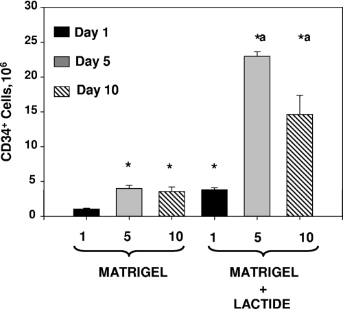
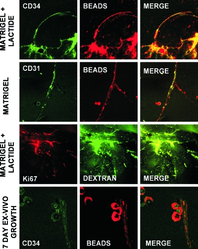
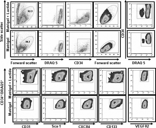
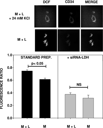
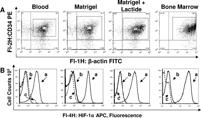
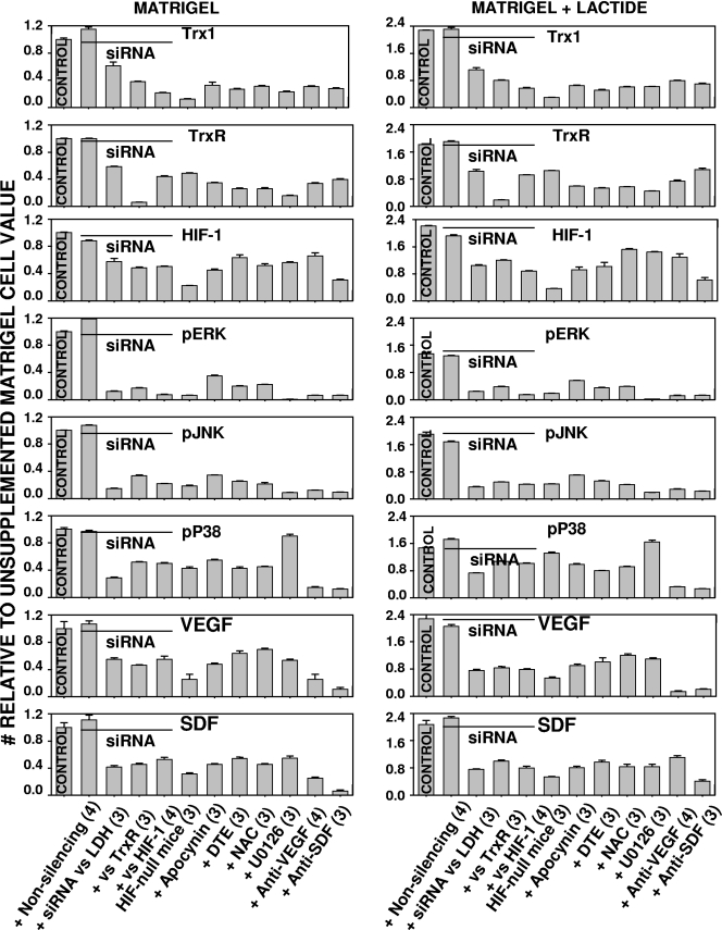
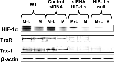
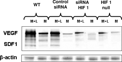
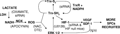
The recruitment and differentiation of circulating stem/progenitor cells (SPCs) in subcutaneous Matrigel in mice was assessed. There were over one million CD34(+) SPCs per Matrigel plug 18 h after Matrigel implantation, and including a polymer to elevate the lactate concentration increased the number of SPCs by 3.6-fold. Intricate CD34(+) cell-lined channels were linked to the systemic circulation, and lactate accelerated cell differentiation as evaluated based on surface marker expression and cell cycle entry. CD34(+) SPCs from lactate-supplemented Matrigel exhibited significantly higher concentrations of thioredoxin 1 (Trx1) and hypoxia-inducible factor 1 (HIF-1) than cells from unsupplemented Matrigel, whereas Trx1 and HIF-1 in CD45(+) leukocytes were not elevated by lactate. Results obtained using small inhibitory RNA (siRNA) specific to HIF-1 and mice with conditionally HIF-1 null myeloid cells indicated that SPC recruitment and lactate-mediated effects were dependent on HIF-1. Cells from lactate-supplemented Matrigel had higher concentrations of phosphorylated extracellular signal-regulated kinases 1 and 2, Trx1, Trx reductase (TrxR), vascular endothelial growth factor (VEGF), and stromal cell-derived factor 1 (SDF-1) than cells from unsupplemented Matrigel. SPC recruitment and protein changes were inhibited by siRNA specific to lactate dehydrogenase, TrxR, or HIF-1 and by oxamate, apocynin, U0126, N-acetylcysteine, dithioerythritol, and antibodies to VEGF or SDF-1. Oxidative stress from lactate metabolism by SPCs accelerated further SPC recruitment and differentiation through Trx1-mediated elevations in HIF-1 levels and the subsequent synthesis of HIF-1-dependent growth factors.
Figures
Similar articles
-
Hyperbaric oxygen stimulates vasculogenic stem cell growth and differentiation in vivo.J Appl Physiol (1985). 2009 Feb;106(2):711-28. doi: 10.1152/japplphysiol.91054.2008. Epub 2008 Nov 20. J Appl Physiol (1985). 2009. PMID: 19023021 Free PMC article.
-
FGF2-dependent neovascularization of subcutaneous Matrigel plugs is initiated by bone marrow-derived pericytes and macrophages.Development. 2008 Feb;135(3):523-32. doi: 10.1242/dev.002071. Epub 2008 Jan 2. Development. 2008. PMID: 18171688
-
Dimethyloxalyl Glycine Regulates the HIF-1 Signaling Pathway in Mesenchymal Stem Cells.Stem Cell Rev Rep. 2020 Aug;16(4):702-710. doi: 10.1007/s12015-019-09947-7. Stem Cell Rev Rep. 2020. PMID: 32372246
-
Transcriptional activation of hypoxia-inducible factor-1 (HIF-1) in myeloid cells promotes angiogenesis through VEGF and S100A8.Proc Natl Acad Sci U S A. 2014 Feb 18;111(7):2698-703. doi: 10.1073/pnas.1320243111. Epub 2014 Feb 4. Proc Natl Acad Sci U S A. 2014. PMID: 24497508 Free PMC article.
-
Lactate, with oxygen, incites angiogenesis.Adv Exp Med Biol. 2008;614:73-80. doi: 10.1007/978-0-387-74911-2_9. Adv Exp Med Biol. 2008. PMID: 18290316 Review.
Cited by
-
Prognostic Significance of Serum Lactic Acid, Lactate Dehydrogenase, and Albumin Levels in Patients with Metastatic Colorectal Cancer.Biomed Res Int. 2018 Dec 9;2018:1804086. doi: 10.1155/2018/1804086. eCollection 2018. Biomed Res Int. 2018. PMID: 30627541 Free PMC article. Clinical Trial.
-
Hyperbaric Oxygen Improves the Survival and Angiogenesis of Fat Grafts after Autologous Fat Transplantation.Biomed Res Int. 2022 May 20;2022:6738959. doi: 10.1155/2022/6738959. eCollection 2022. Biomed Res Int. 2022. Retraction in: Biomed Res Int. 2023 Dec 29;2023:9834539. doi: 10.1155/2023/9834539. PMID: 35647192 Free PMC article. Retracted.
-
Oxidative stress is fundamental to hyperbaric oxygen therapy.J Appl Physiol (1985). 2009 Mar;106(3):988-95. doi: 10.1152/japplphysiol.91004.2008. Epub 2008 Oct 9. J Appl Physiol (1985). 2009. PMID: 18845776 Free PMC article. Review.
-
Wnt signaling directs a metabolic program of glycolysis and angiogenesis in colon cancer.EMBO J. 2014 Jul 1;33(13):1454-73. doi: 10.15252/embj.201488598. Epub 2014 May 13. EMBO J. 2014. PMID: 24825347 Free PMC article.
-
TREM2-mediated Macrophage Glycolysis Promotes Skin Wound Angiogenesis via the Akt/mTOR/HIF-1α Signaling Axis.Curr Med Sci. 2024 Dec;44(6):1280-1292. doi: 10.1007/s11596-024-2946-3. Epub 2024 Dec 14. Curr Med Sci. 2024. PMID: 39672999
References
-
- Ali, M., F. Yasui, S. Matsugo, and T. Konishi. 2000. The lactate-dependent enhancement of hydroxyl radical generation by the Fenton reaction. Free Radic. Res. 32429-438. - PubMed
-
- Andoh, T., C. Chiueh, and P. Chock. 2003. Cyclic GMP-dependent protein kinase regulates the expression of thioredoxin and thioredoxin peroxidase-1 during hormesis in response to oxidative stress-induced apoptosis. J. Biol. Chem. 278885-890. - PubMed
-
- Arai, R., H. Masutani, J. Yodoi, V. Debbas, F. Laurindo, A. Stern, and H. Monteiro. 2006. Nitric oxide induces thioredoxin-1 nuclear translocation: possible association with the p21Ras survival pathway. Biochem. Biophys. Res. Commun. 3481254-1260. - PubMed
-
- Asahara, T., T. Murohara, A. Sullivan, M. Silver, R. van der Zee, T. Li, B. Witzenbichler, G. Schatteman, and J. M. Isner. 1997. Isolation of putative progenitor endothelial cells for angiogenesis. Science 275964-967. - PubMed
-
- Balasubramaniam, V., C. F. Mervis, A. M. Maxey, N. E. Markham, and S. H. Abman. 2007. Hyperoxia reduces bone marrow, circulating, and lung endothelial progenitor cells in the developing lung: implications for the pathogenesis of bronchopulmonary dysplasia. Am. J. Physiol. Lung Cell. Mol. Physiol. 292L1073-L1084. - PubMed
Publication types
MeSH terms
Substances
Grants and funding
LinkOut - more resources
Full Text Sources
Other Literature Sources
Medical
Molecular Biology Databases
Research Materials
Miscellaneous