. 2008 Sep;173(3):792-802.
doi: 10.2353/ajpath.2008.080259. Epub 2008 Aug 18.

Long-term survival of transplanted stem cells in immunocompetent mice with muscular dystrophy

Affiliations

Long-term survival of transplanted stem cells in immunocompetent mice with muscular dystrophy

Gregory Q Wallace et al. Am J Pathol. 2008 Sep.

Abstract

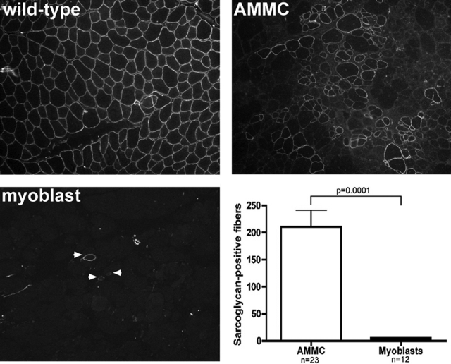
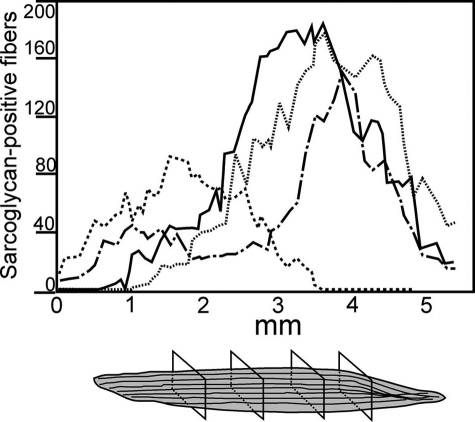
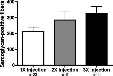
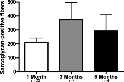
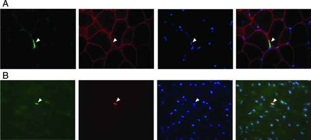
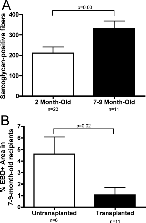
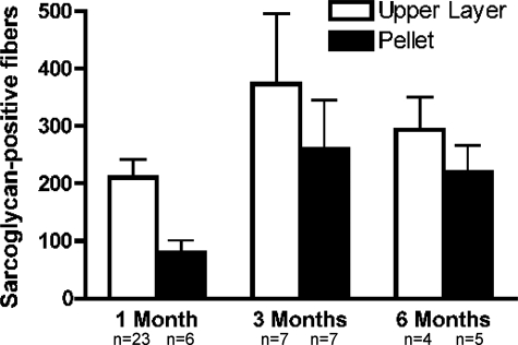
Satellite cells refer to resident stem cells in muscle that are activated in response to damage or disease for the regeneration and repair of muscle fibers. The use of stem cell transplantation to treat muscular diseases has been limited by impaired donor cell survival attributed to rejection and an unavailable stem cell niche. We isolated a population of adult muscle mononuclear cells (AMMCs) from normal, strain-matched muscle and transplanted these cells into delta-sarcoglycan-null dystrophic mice. Distinct from other transplant studies, the recipient mice were immunocompetent with an intact endogenous satellite cell pool. We found that AMMCs were 35 times more efficient at restoring sarcoglycan compared with cultured myoblasts. Unlike cultured myoblasts, AMMC-derived muscle fibers expressed sarcoglycan protein throughout their entire length, consistent with enhanced migratory ability. We examined the capacity of single injections of AMMCs to provide long-term benefit for muscular dystrophy and found persistent regeneration after 6 months, consistent with augmentation of the endogenous stem cell pool. Interestingly, AMMCs were more effectively engrafted into aged dystrophic mice for the regeneration of large clusters of sarcoglycan-positive muscle fibers, which were protected from damage, suggesting that the stem cell niche in older muscle remains permissive.


Figures

Figure 1
Figure 1
The myogenic ability of AMMCs is greater than primary myoblast cultures. Fluorescent staining for γ-sarcoglycan protein revealed large clusters of donor-derived myofibers 1 month after transplantation in Sgcd-null muscles injected once with 105 AMMCs. In contrast, injection of cultured myoblasts resulted in scattered, isolated sarcoglycan-positive muscle fibers. Muscles injected with AMMCs had 35-fold more sarcoglycan-positive myofibers than those injected with cultured myoblasts. Sarcoglycan-positive fibers were scored on at least 140 sections per recipient muscle. Shown are the average values from the section with the most sarcoglycan-positive fibers from each recipient. Original magnifications, ×100.
Figure 2
Figure 2
AMMCs restore sarcoglycan protein throughout the length of transplanted muscles. Sarcoglycan-positive myofibers were scored by immunofluorescence microscopy in at least 140 serial sections throughout 5 mm of recipient tibialis anterior muscles. Shown are data from four representative muscles from Sgcd-null mice injected once with 105 AMMCs and analyzed 1 month after transplantation.
Figure 3
Figure 3
AMMC-derived muscle fibers resist degeneration. For each recipient, the serial tibialis anterior section containing the most donor-derived myofibers was stained with anti-γ-sarcoglycan antibodies (green) and EBD (red), which binds albumin and enters damaged muscle fibers. Nuclei are stained with DAPI (blue). Damaged muscle fibers were detected throughout the muscle, but none of the 3711 donor-derived myofibers observed in 33 recipient muscles contained EBD. Original magnifications, ×100.
Figure 4
Figure 4
Transplantation of AMMCs reduces exercise-induced damage in biceps brachii. The percentage of cross-sectional area that contained EBD (EBD)-positive fibers was determined in untransplanted biceps brachii and muscles injected twice with 105 AMMCs. Exercised mice were subjected to three sessions of downhill running to induce damage in transplanted and untransplanted biceps muscles. Muscle damage was almost 50% lower in mice that received injections of AMMCs. P = 0.05 for untransplanted, exercised animals compared to transplanted, exercised animals.
Figure 5
Figure 5
Multiple injections of AMMCs increase sarcoglycan expression. Sarcoglycan-positive fibers were quantified in tibialis anterior muscles 1 month after receiving the first of one, two, or three injections of 105 AMMCs. Three injections increased the number of sarcoglycan-positive fibers (P = 0.04, one injection compared to three injections) indicating that increased exposure to donor-derived cells and sarcoglycan expression did not adversely affect engraftment and survival.
Figure 6
Figure 6
Long-term survival of donor-derived fibers in AMMC-transplanted muscles. Tibialis anterior muscles of mice given single injections of AMMCs were examined by immunofluorescence microscopy 1, 3, or 6 months after injection. Sarcoglycan-positive fibers were maintained 3 and 6 months after the single injection of donor cells. Because multiple rounds of regeneration occur during this period, this finding is consistent with successful replacement of the satellite cell pool and protection against the disease process for those fibers that express sarcoglycan.
Figure 7
Figure 7
Pax7-positive donor-derived AMMCs localize in a satellite cell position. Sgcd-null mice were injected with GFP-expressing donor cells. A: AMMCs from a transgenic GFP mouse were injected into a dystrophic recipient Sgcd-null mouse and were detected 1 month later closely associated with the extracellular sarcolemma (arrowhead), consistent with the localization of satellite cells. GFP-AMMCs were identified by GFP (green) and nuclear staining (DAPI, blue) and the muscle plasma membrane was labeled with anti-dystrophin antibodies (red). B: Pax7 (red) was also detected in GFP-AMMCs residing near the sarcolemma (arrowhead). Original magnifications, ×630.
Figure 8
Figure 8
Transplantation of AMMCs is more effective in aged Sgcd-null mice recipients. A: Sarcoglycan restoration was compared in 1.5- to 2.5-month-old recipients and 7- to 10-month-old recipients of AMMC transplants. Older mice receiving AMMCs had 60% more sarcoglycan-positive fibers than younger recipients (P = 0.03). B: Tibialis anterior sections of transplanted and untransplanted 7- to 10-month-old recipient Sgcd-null mice were stained with EBD to measure muscle damage. Aged mice receiving AMMC transplants had fewer dye-positive fibers than age-matched, untransplanted Sgcd-null mice (P = 0.02).
Figure 9
Figure 9
Fractionation of AMMCs through a Ficoll gradient. One month after transplantation, AMMCs resulted in 2.5× more donor-derived muscle fibers compared to Ficoll pellet transplants. At 3 and 6 months after transplantation, however, sarcoglycan-positive fibers were nearly equal in recipients of AMMCs and Ficoll pellet cells. The pellet fraction had increased expression of laminin α2, integrin α5, Pax7, and Pax3 suggesting that fragments of the stem cell niche may remain intact and promote long-term regeneration.

References

    1. Mauro A. Satellite cell of skeletal muscle fibers. J Biophys Biochem Cytol. 1961;9:493–495. - PMC - PubMed
    1. Moss FP, Leblond CP. Satellite cells as the source of nuclei in muscles of growing rats. Anat Rec. 1971;170:421–435. - PubMed
    1. Morgan JE, Watt DJ, Sloper JC, Partridge TA. Partial correction of an inherited biochemical defect of skeletal muscle by grafts of normal muscle precursor cells. J Neurol Sci. 1988;86:137–147. - PubMed
    1. Morgan JE. Practical aspects of myoblast implantation: investigations on two inherited myopathies in animals. Adv Exp Med Biol. 1990;280:86–95. - PubMed
    1. Karpati G, Pouliot Y, Zubrzycka-Gaarn E, Carpenter S, Ray PN, Worton RG, Holland P. Dystrophin is expressed in mdx skeletal muscle fibers after normal myoblast implantation. Am J Pathol. 1989;135:27–32. - PMC - PubMed

Publication types

MeSH terms