. 2008 Aug 26;105(34):12445-50.
doi: 10.1073/pnas.0804186105. Epub 2008 Aug 18.

Oncogenic bystander radiation effects in Patched heterozygous mouse cerebellum

Affiliations

Oncogenic bystander radiation effects in Patched heterozygous mouse cerebellum

Mariateresa Mancuso et al. Proc Natl Acad Sci U S A. .

Abstract

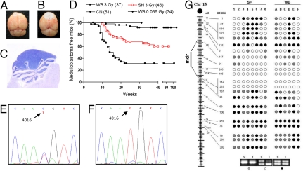
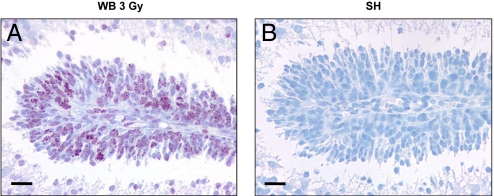
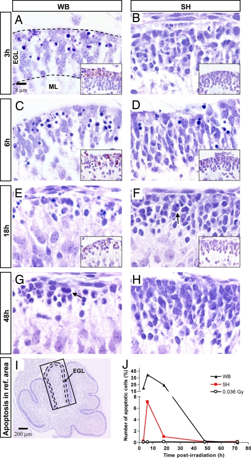
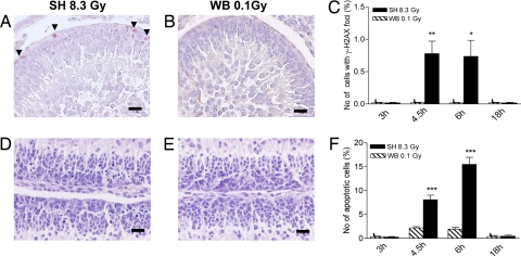
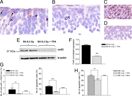
The central dogma of radiation biology, that biological effects of ionizing radiation are a direct consequence of DNA damage occurring in irradiated cells, has been challenged by observations that genetic/epigenetic changes occur in unexposed "bystander cells" neighboring directly-hit cells, due to cell-to-cell communication or soluble factors released by irradiated cells. To date, the vast majority of these effects are described in cell-culture systems, while in vivo validation and assessment of biological consequences within an organism remain uncertain. Here, we describe the neonatal mouse cerebellum as an accurate in vivo model to detect, quantify, and mechanistically dissect radiation-bystander responses. DNA double-strand breaks and apoptotic cell death were induced in bystander cerebellum in vivo. Accompanying these genetic events, we report bystander-related tumor induction in cerebellum of radiosensitive Patched-1 (Ptch1) heterozygous mice after x-ray exposure of the remainder of the body. We further show that genetic damage is a critical component of in vivo oncogenic bystander responses, and provide evidence supporting the role of gap-junctional intercellular communication (GJIC) in transmission of bystander signals in the central nervous system (CNS). These results represent the first proof-of-principle that bystander effects are factual in vivo events with carcinogenic potential, and implicate the need for re-evaluation of approaches currently used to estimate radiation-associated health risks.


Conflict of interest statement

The authors declare no conflict of interest.

Figures

Fig. 1.
Fig. 1.
Irradiation set up for shielded irradiation. (A) Neonatal Ptch1+/− and Ptch1+/+ mice placed in polystyrene boxes were irradiated with heads and upper body shielded by individual custom-built lead cylinders. (B) Demarcation between exposed and shielded regions at P10 due to hair-growth delay in exposed skin. (C) Characteristics of the lead shields.
Fig. 2.
Fig. 2.
Medulloblastoma development in Ptch1+/− mice. (A) Normal brain. (B) Macroscopic aspect of cerebellar tumors developing in the posterior fossa. (C) Histology of medulloblastoma, composed of tightly packed, small round cells with minimal surrounding cytoplasm. (D) Kaplan–Meier kinetic analysis of medulloblastoma in whole-body irradiated (WB), shield-irradiated (SH), and control Ptch1+/− mice (CN). Included is the tumor-free survival at 31 weeks of mice that were WB irradiated with a dose of 0.036 Gy (WB-0.036), equivalent to the scatter dose to SH cerebellum. (E and F) Representative electropherogram of genomic and tumor DNAs showing retention of both Ptch1 alleles in normal tissue (E) and loss of WT Ptch1 allele in medulloblastomas (F). (G) Analysis of chr-13 LOH in tumors from WB and SH Ptch1+/− mice. The distance of microsatellite markers (D13Mit) from the centromere is given in cM. Closed circles indicate no LOH; open circles denote LOH; gray circles indicate not informative (NI) markers; mdr: minimum deleted region.
Fig. 3.
Fig. 3.
γ-H2AX immunohistochemical staining of the EGL of P2 cerebellum after irradiation. (A) A large fraction (> 85%) of GCPs exhibited numerous nuclear foci of γ-H2AX at 0.5 h post-3Gy whole-body (WB) irradiation. (B) Total lack of γ-H2AX staining in the EGL of shielded mice (SH) at 0.5 h postirradiation. (Scale bars, 20 μm.)
Fig. 4.
Fig. 4.
Levels and kinetics of apoptosis in exposed and shielded P2 mouse cerebellum. (A–J) Quantification of apoptosis in the anterodorsal cardinal lobe (boxed in I). (A–D Apoptosis at 3 and 6 h postirradiation in the EGL of WB- relative to SH-exposed mice. (A–F, Insets) Immunohistochemical analysis of caspase-3 activation. (E and G) Thinning of the EGL in WB-exposed and to lower degree in SH-exposed mice (F). Cell repopulation with mitotic figures in WB-exposed mice at 48 h (G). Compensatory hyperplasia at 48 h in SH-exposed EGL (F and H). Black dashes in A and I delineate the EGL. (J) Percentage of apoptotic cells as a function of time postirradiation in WB- and SH-exposed mice.
Fig. 5.
Fig. 5.
Radiation damage by expected scatter dose in exposed vs. bystander EGL. (A and B) γ-H2AX positivity in the outer EGL of SH-8.3 Gy mice at 6h postirradiation compared with undetectable staining after exposure to the scatter dose (0.1 Gy). (D and E) Increased apoptosis in EGL of SH-8.3Gy mice at 6 h postirradiation compared with very rare apoptosis after a 0.1 Gy dose. (C and F) Percentage of γ-H2AX-positive and apoptotic cells in cerebellum at 3, 4.5, 6, and 18 h post-8.3-Gy (SH) and 0.1 Gy (WB) irradiation. *, P = 0.0139; **, P = 0.0015; ***, P = 0.0001. (Scale bars, 20 μm.)
Fig. 6.
Fig. 6.
Inhibition of GJICs reverses bystander effects in cerebellum. (A) Immunohistochemical analysis showing widespread expression of Cx43 protein in the EGL at 6h post-8.3Gy SH irradiation. Arrows: Cx43 expression at cell–cell contacts. (B) Lack of Cx43 positivity in cerebellum of mice receiving combined TPA and SH-8.3Gy treatment. (C) Strong Cx43 expression in the postnatal SVZ, and (D) reduced expression in the SVZ of SH-8.3Gy mice injected with TPA are shown for comparison. (E and F) Western blot analysis showing decrease of Cx43 expression in TPA-treated relative to -untreated mice (P = 0.0025). (G) TPA (gray columns) dramatically reduced the levels of γ-H2AX-positive (P = 0.0075) and apoptotic cells (P < 0.0001) in shielded cerebellum; note numerous apoptotic figures in A, compared with sporadic apoptosis (white arrowhead) in B. No significant effect of nimesulide (Nms; white columns) administration on levels of short-term cellular responses. (H) TPA, but not Nms, significantly reduced apoptosis in cerebellum directly exposed to 3 Gy.

References

    1. Little JB. Radiation carcinogenesis. Carcinogenesis. 2000;21:397–404. - PubMed
    1. Sowa M, Arthurs BJ, Estes BJ, Morgan WF. Effects of ionizing radiation on cellular structures, induced instability and carcinogenesis. EXS. 2006;96:293–301. - PubMed
    1. Nagasawa H, Little JB. Induction of sister chromatid exchanges by extremely low doses of alpha-particles. Cancer Res. 1992;52:6394–6396. - PubMed
    1. Azzam EI, de Toledo SM, Gooding T, Little JB. Intercellular communication is involved in the bystander regulation of gene expression in human cells exposed to very low fluences of alpha particles. Radiat Res. 1998;150:497–504. - PubMed
    1. Huang L, Kim PM, Nickoloff JA, Morgan WF. Targeted and nontargeted effects of low-dose ionizing radiation on delayed genomic instability in human cells. Cancer Res. 2007;67:1099–1104. - PubMed

Publication types

LinkOut - more resources