

Genetic disruption of cyclooxygenase-2 does not improve histological or behavioral outcome after traumatic brain injury in mice
- PMID: 18711748
- PMCID: PMC2737709
- DOI: 10.1002/jnr.21809
Genetic disruption of cyclooxygenase-2 does not improve histological or behavioral outcome after traumatic brain injury in mice
Abstract
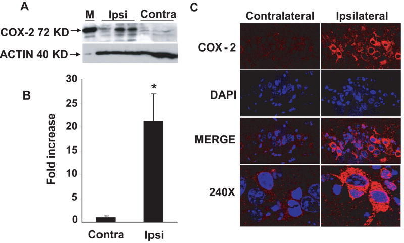
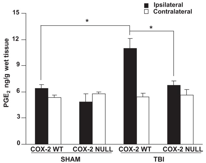
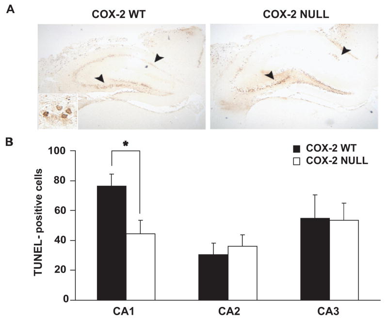
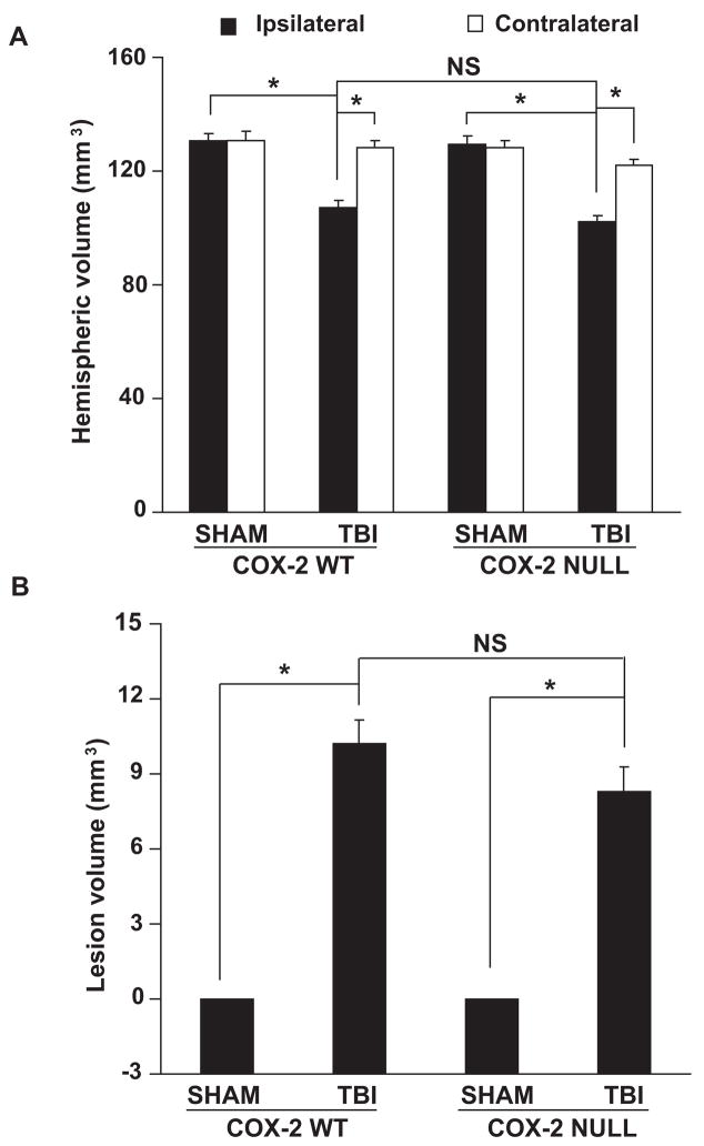
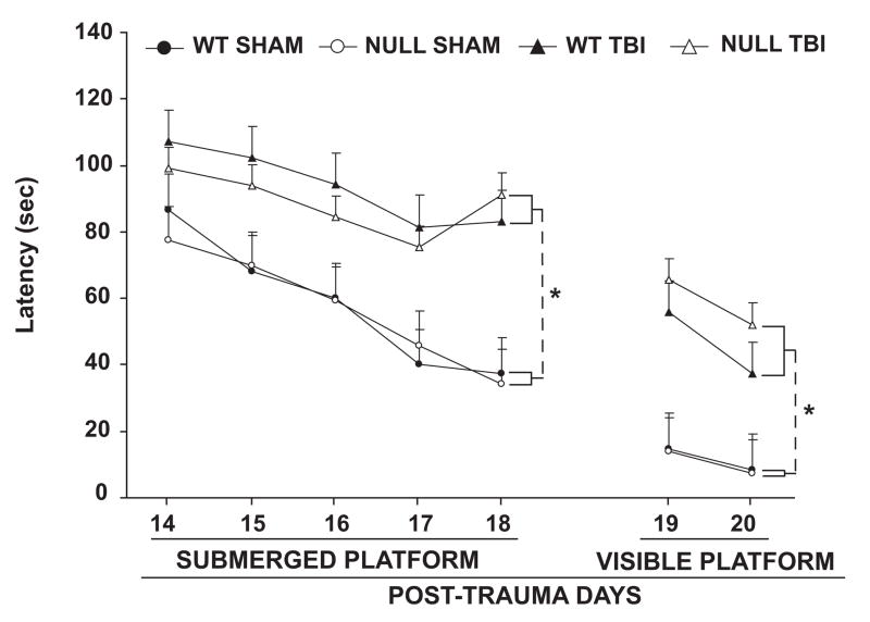
Increasing evidence suggests a role for cyclooxygenase-2 (COX-2) in traumatic brain injury (TBI). In the present study, the role of COX-2 in TBI was investigated using COX-2 gene-disrupted (COX-2 null) mice and wild-type (WT) controls that were subjected to the controlled cortical impact (CCI) model of TBI. There was increased expression of COX-2 in ipsilateral hippocampus in WT mice subjected to CCI. CCI resulted in a significant increase in prostaglandin E(2) concentrations in WT compared with COX-2 null hippocampi. There was a significant increase in TUNEL staining of CA1 neurons 24 hr after CCI in WT, but not in COX-2 null mice, compared with sham-operated controls, which is consistent with a protective role for COX-2 in the early phase of injury after TBI. However, there was no difference in lesion volume 21 days after CCI in COX-2 null and WT mice. COX-2 gene disruption did not alter Morris water maze performance. Taken together, these results suggest only a minor role for COX-2 activity in determining outcome after TBI in mouse.
(c) 2008 Wiley-Liss, Inc.
Figures
References
-
- Alkayed NJ, Birks EK, Hudetz AG, Roman RJ, Henderson L, Harder DR. Inhibition of brain P-450 arachidonic acid epoxygenase decreases baseline cerebral blood flow. Am J Physiol. 1996;271(4 Pt 2):H1541–1546. - PubMed
-
- Alloza I, Baxter A, Chen Q, Matthiesen R, Vandenbroeck K. Celecoxib inhibits interleukin-12 alphabeta and beta2 folding and secretion by a novel COX2-independent mechanism involving chaperones of the endoplasmic reticulum. Mol Pharmacol. 2006;69(5):1579–1587. - PubMed
-
- Belton O, Fitzgerald DJ. Cyclooxygenase isoforms and atherosclerosis. Expert Rev Mol Med. 2003;5(9):1–18. - PubMed
-
- Cernak I, O’Connor C, Vink R. Inhibition of cyclooxygenase 2 by nimesulide improves cognitive outcome more than motor outcome following diffuse traumatic brain injury in rats. Exp Brain Res. 2002;147(2):193–199. - PubMed
-
- Chang JW, Coleman PD, O’Banion MK. Prostaglandin G/H synthase-2 (cyclooxygenase-2) mRNA expression is decreased in Alzheimer’s disease. Neurobiology of aging. 1996;17(5):801–808. - PubMed
Publication types
MeSH terms
Substances
Grants and funding
LinkOut - more resources
Full Text Sources
Medical
Research Materials
Miscellaneous
