. 2009 Mar;457(5):989-1001.
doi: 10.1007/s00424-008-0574-6. Epub 2008 Aug 19.

Muscle wasting and interleukin-6-induced atrogin-I expression in the cachectic Apc ( Min/+ ) mouse

Affiliations

Muscle wasting and interleukin-6-induced atrogin-I expression in the cachectic Apc ( Min/+ ) mouse

Kristen A Baltgalvis et al. Pflugers Arch. 2009 Mar.

Abstract

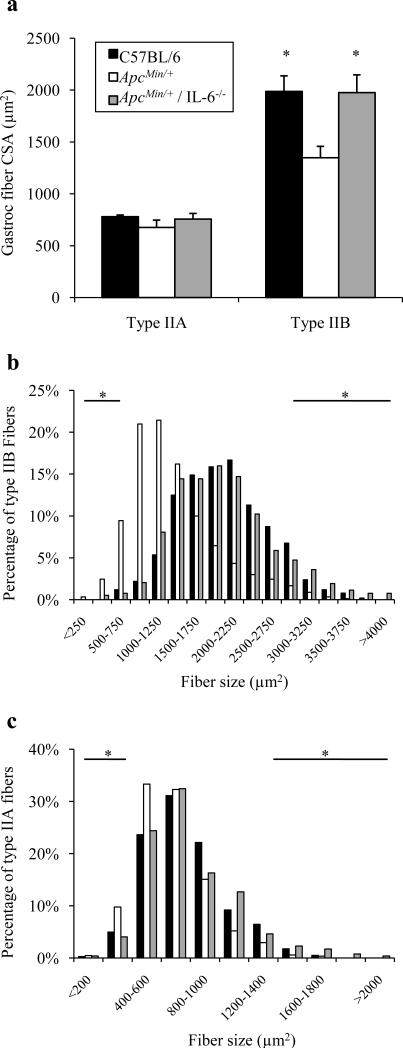
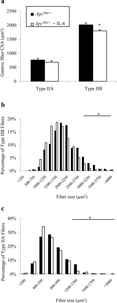
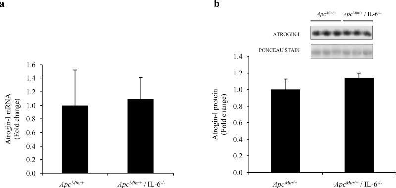
Interleukin-6 (IL-6) is necessary for cachexia in Apc ( Min/+ ) mice, but the mechanisms inducing this myofiber wasting have not been established. The purpose of this study was to examine gastrocnemius muscle wasting in the Apc ( Min/+ ) mouse and to determine IL-6 regulated mechanisms contributing to muscle loss. Gastrocnemius type IIB mean fiber cross-sectional area (CSA) from Apc ( Min/+ ) mice decreased 32% between 13 and 22 weeks of age. Apc ( Min/+ ) mice lacking IL-6 did not have type IIB fiber atrophy, while overexpression of circulating IL-6 exacerbated the loss of type IIB fiber CSA in Apc ( Min/+ ) mice. Muscle Atrogin-I mRNA expression was induced at least ninefold at 18 and 22 weeks of age compared to 13-week-old mice. Atrogin-I gene expression was also induced by overexpression of circulating IL-6. These data suggest that high circulating IL-6 levels induce type IIB fiber CSA loss in Apc ( Min/+ ) mice, and circulating IL-6 is sufficient to regulate Atrogin-I gene expression in cachectic mice.


Figures

Fig. 1
Fig. 1
Time course of gastrocnemius muscle wasting in ApcMin/+ mice. Gastrocnemius muscle mass (a), mean gastrocnemius fiber CSA of type IIA and type IIB fibers (b), frequency distribution for type IIB gastrocnemius fibers (c), and frequency distribution of type IIA fibers (d). Muscles were harvested from 13-wk-old (n=13), 18-wk-old (n=11), and 22-wk-old (n=7) ApcMin/+ mice. Values are means ± SE. *Signifies different from 13-wk-old mice.
Fig. 2
Fig. 2
Gene and protein expression related to protein degradation in cachectic ApcMin/+ mouse gastrocnemius muscle. IL-6 mRNA (a) and STAT-3 phosphorylation (b) were indicators of IL-6 signaling. MuRF-I mRNA (c) and Atrogin-I mRNA (d) were measured in 13-, 18-, and 22-wk-old ApcMin/+ mice. Atrogin-I protein levels were also elevated during cachexia (e). There was a significant correlation (r=-0.675; P=0.002) between Atrogin-I mRNA and gastrocnemius muscle mass (f). Values are means ± SE. *Signifies different from 13-wk-old mice.
Fig. 3
Fig. 3
Gastrocnemius fiber CSA in 26-week-old ApcMin/+ mice lacking IL-6. Mean gastrocnemius type IIA and type IIB fiber CSA (a) in 26-week old C57BL/6, ApcMin/+, and ApcMin/+ / IL-6-/- mice. Type IIB (b) and type IIA (c) fiber frequency distributions in C57BL/6, ApcMin/+, and ApcMin/+ / IL-6-/- mice. Values are means ± SE. *Signifies both C57BL/6 and ApcMin/+ / IL-6-/- different from ApcMin/+ mice.
Fig. 4
Fig. 4
Over-expression of circulating IL-6 in ApcMin/+ mice. Mean gastrocnemius type IIA and type IIB fiber CSA (a) and fiber frequency distributions of type IIB (b) and type IIA fibers (c) following 4 weeks of increased IL-6. Values are means ± SE. *Signifies different from ApcMin/+ Control.
Fig. 5
Fig. 5
Atrogin-I and MuRF-I mRNA and protein expression in gastrocnemius muscle from mice over-expressing circulating IL-6. Atrogin-I and MuRF-I gene expression during increased circulating IL-6 in cachectic ApcMin/+ mice (a). There was a positive correlation between Atrogin-I mRNA levels and plasma IL-6 in ApcMin/+ mice (b). Atrogin-I protein levels during IL-6 over-expression in ApcMin/+ mice (c). Atrogin-I and MuRF-I gene expression during increased circulating IL-6 in non-cachectic, wild-type mice (d). Atrogin-I protein levels during IL-6 over-expression in C57BL/6 mice (e). Values are means ± SE. *Signifies different from Control.
Fig. 6
Fig. 6
Atrogin-I mRNA and protein expression in 6-mo-old ApcMin/+ and ApcMin/+ / IL-6-/- mice. Atrogin-I mRNA (a) and Atrogin-I protein (b) in 6-mo-old cachectic (ApcMin/+) and non-cachectic (ApcMin/+ / IL-6-/-) mice. Values are means ± SE.

References

    1. Price SA, Tisdale MJ. Mechanism of inhibition of a tumor lipid-mobilizing factor by eicosapentaenoic acid. Cancer Res. 1998;58:4827–4831. - PubMed
    1. Giordano A, Calvani M, Petillo O, Carteni M, Melone MR, Peluso G. Skeletal muscle metabolism in physiology and in cancer disease. J Cell Biochem. 2003;90:170–186. - PubMed
    1. al-Majid S, McCarthy DO. Resistance exercise training attenuates wasting of the extensor digitorum longus muscle in mice bearing the colon-26 adenocarcinoma. Biol Res Nurs. 2001;2:155–166. - PubMed
    1. Ardies CM. Exercise, cachexia, and cancer therapy: a molecular rationale. Nutr Cancer. 2002;42:143–157. - PubMed
    1. Khalfoun B, Thibault F, Watier H, Bardos P, Lebranchu Y. Docosahexaenoic and eicosapentaenoic acids inhibit in vitro human endothelial cell production of interleukin-6. Adv Exp Med Biol. 1997;400B:589–597. - PubMed

Publication types

MeSH terms

LinkOut - more resources