Review
. 2008 Nov 1;123(9):1979-90.
doi: 10.1002/ijc.23858.

Optical contrast agents and imaging systems for detection and diagnosis of cancer

Affiliations
Review

Optical contrast agents and imaging systems for detection and diagnosis of cancer

Mark C Pierce et al. Int J Cancer. .

Abstract

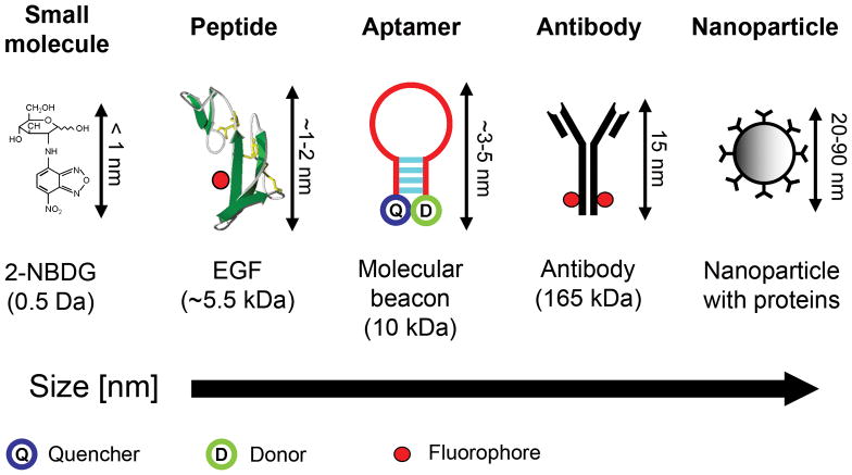
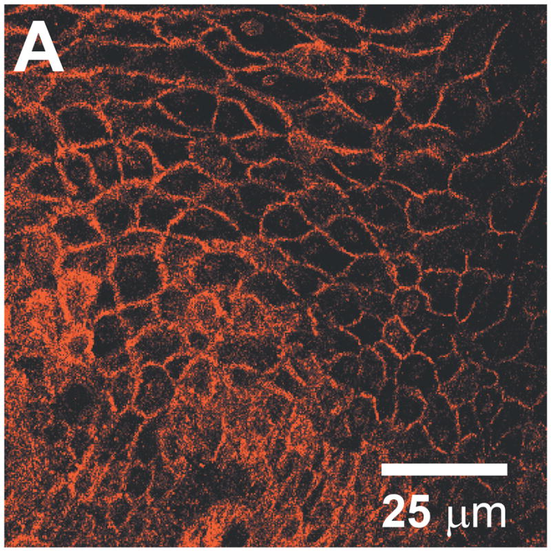
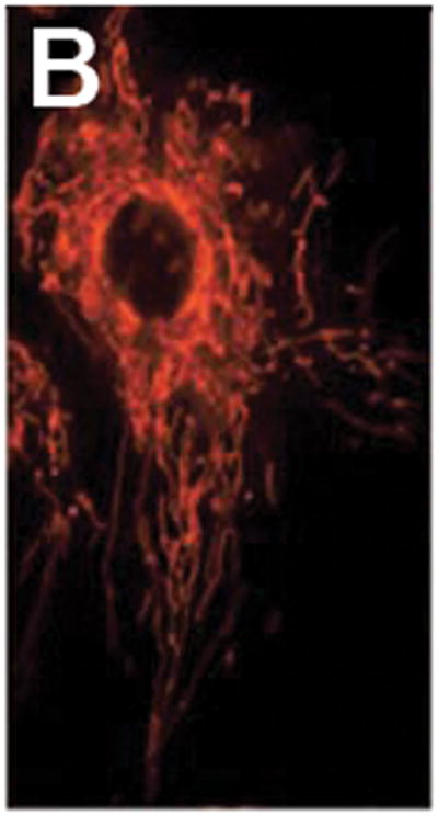
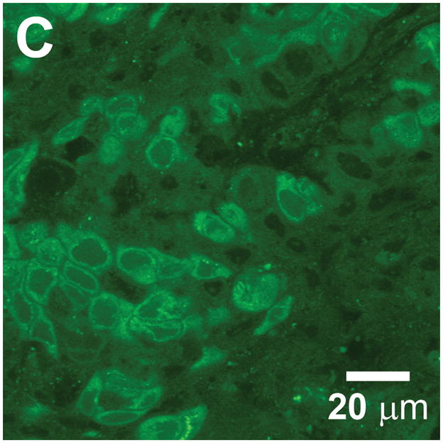
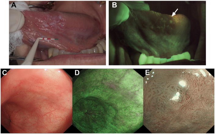
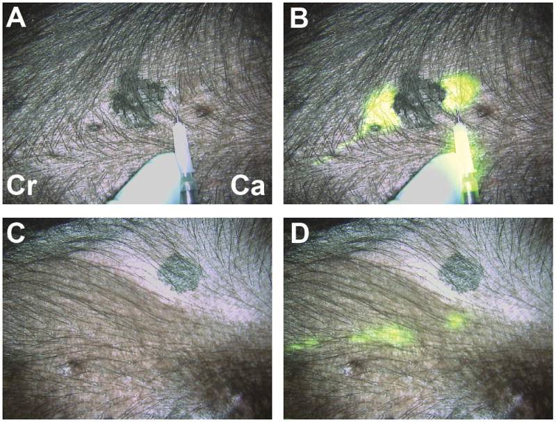
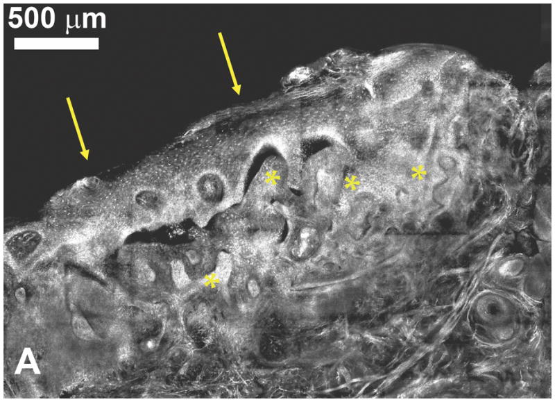
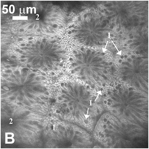
Molecular imaging has rapidly emerged as a discipline with the potential to impact fundamental biomedical research and clinical practice. Within this field, optical imaging offers several unique capabilities, based on the ability of cells and tissues to effect quantifiable changes in the properties of visible and near-infrared light. Beyond endogenous optical properties, the development of molecularly targeted contrast agents enables disease-specific morphologic and biochemical processes to be labeled with unique optical signatures. Optical imaging systems can then provide real-time visualization of pathophysiology at spatial scales from the subcellular to whole organ levels. In this article, we review fundamental techniques and recent developments in optical molecular imaging, emphasizing laboratory and clinical systems that aim to visualize the microscopic and macroscopic hallmarks of cancer.


Figures

Figure 1
Figure 1
Five classes of molecular-specific optical contrast agent. From left to right in order of increasing size: Small molecules including glucose and peptides can be functionalized with fluorescent dyes. Aptamers can be designed to form activatable “smart probes”, with fluorescence quenched until target binding. Antibody probes are generally functionalized with fluorescent dyes in the Fc domain. Targeting molecules can be coupled to nanoparticle-based optical reporters, including gold nanoparticles and quantum dots.
Figure 2
Figure 2
Cell and tissue labeling with molecular-specific optical contrast agents. (A) Confocal reflectance image of an abnormal cervical biopsy section labeled with anti-EGFR gold conjugates, illuminated at 647 nm. Scale bar ~ 25 μm. Reproduced with permission from [18]. (B) Epi-fluorescence image of cultured HDF cells labeled with dual FRET-based molecular beacons, designed to exhibit specific binding to target K-ras mRNA. Reproduced with permission from [24]. (C) Confocal fluorescence image of NBDG labeled human oral biopsy section, demonstrating intracellular uptake of contrast agent via glucose transporters. Reproduced with permission from [26].
Figure 2
Figure 2
Cell and tissue labeling with molecular-specific optical contrast agents. (A) Confocal reflectance image of an abnormal cervical biopsy section labeled with anti-EGFR gold conjugates, illuminated at 647 nm. Scale bar ~ 25 μm. Reproduced with permission from [18]. (B) Epi-fluorescence image of cultured HDF cells labeled with dual FRET-based molecular beacons, designed to exhibit specific binding to target K-ras mRNA. Reproduced with permission from [24]. (C) Confocal fluorescence image of NBDG labeled human oral biopsy section, demonstrating intracellular uptake of contrast agent via glucose transporters. Reproduced with permission from [26].
Figure 3
Figure 3
(A) White light image of the ventral tongue of a patient with an oral premalignant lesion, which when biopsied was confirmed to be severe dysplasia. (B) Autofluorescence image with the region of fluorescence visualization loss and biopsy location indicated by the arrow. Reproduced with permission from [41]. (C) During initial inspection of a region of Barrett’s esophagus with high-resolution white-light endoscopy, no abnormalities were seen. (D) On inspection with AFI, a small area with suspicious purple autofluorescence was seen at the 1 o’clock position. (E) Subsequent detailed inspection by narrowband imaging showed irregular mucosal and vascular patterns and presence of abnormal blood vessels. Histology from targeted biopsies showed adenocarcinoma. Reproduced with permission from [43].
Figure 4
Figure 4
Real-time intraoperative near-infrared fluorescent sentinel lymph node (SLN) mapping. (A,B) At T = 0, four peri-tumoral, subcutaneous injections of 1 nmol of HSA800 were made around a primary melanoma on the ventral left torso. Two dominant lymphatic channels, one cranial (Cr) and one caudal (Ca) were found. (C, D) The caudal channel was followed until two SLNs were identified at T = 15 secs. Images shown include color video (A, C) and a pseudo-colored (lime green) merge with near-infrared fluorescence (B, D). Reproduced with permission from [44].
Figure 5
Figure 5
(A) Confocal submosaic of a superficial BCC shows 8 × 6 frames stitched together to show an equivalent ~ 4× magnified view. Bright nuclei are more clearly seen in epidermis along the peripheral edge (arrows) and the BCC (*) in the underlying deep dermis. Scale bar = 500 μm. For corresponding histopathology, see [46]. Reproduced with permission from [46]. (B) Single scan confocal images (FOV = 500 μm × 500 μm) collected in vivo using a prototype endoscope design suitable for full colonoscopy. Confocal image of descending colon mucosa. Topical application of acriflavine strongly stained the superficial cells only. 1 = Goblet cells, 2 = Crypt lumen. Reproduced with permission from [49].
Figure 5
Figure 5
(A) Confocal submosaic of a superficial BCC shows 8 × 6 frames stitched together to show an equivalent ~ 4× magnified view. Bright nuclei are more clearly seen in epidermis along the peripheral edge (arrows) and the BCC (*) in the underlying deep dermis. Scale bar = 500 μm. For corresponding histopathology, see [46]. Reproduced with permission from [46]. (B) Single scan confocal images (FOV = 500 μm × 500 μm) collected in vivo using a prototype endoscope design suitable for full colonoscopy. Confocal image of descending colon mucosa. Topical application of acriflavine strongly stained the superficial cells only. 1 = Goblet cells, 2 = Crypt lumen. Reproduced with permission from [49].
Figure 6
Figure 6
(A) E-selectin is constitutively expressed in a subset of vessels expressing PECAM-1. The green channel is obtained by two-photon excitation of FITC–anti-PECAM-1 mAb using a Ti:Al2O3 laser at 800 nm, whereas the red channel is obtained by one-photon excitation of Cy5.5–anti-E-selectin mAb using a HeNe laser at 633 nm. Scale bar = 100 μm. Reproduced with permission from [58]. (B) Adipocytes imaged with coherent anti-Stokes Raman scattering (red) and collagen fibrils imaged with second harmonic generation (green) in a mammary gland. Scale bar = 25 μm. Reproduced with permission from [71].
Figure 6
Figure 6
(A) E-selectin is constitutively expressed in a subset of vessels expressing PECAM-1. The green channel is obtained by two-photon excitation of FITC–anti-PECAM-1 mAb using a Ti:Al2O3 laser at 800 nm, whereas the red channel is obtained by one-photon excitation of Cy5.5–anti-E-selectin mAb using a HeNe laser at 633 nm. Scale bar = 100 μm. Reproduced with permission from [58]. (B) Adipocytes imaged with coherent anti-Stokes Raman scattering (red) and collagen fibrils imaged with second harmonic generation (green) in a mammary gland. Scale bar = 25 μm. Reproduced with permission from [71].

References

    1. Weissleder R, Pittet MJ. Imaging in the era of molecular oncology. Nature. 2008;452:580–589. - PMC - PubMed
    1. Massoud TF, Gambhir SS. Integrating noninvasive molecular imaging into molecular medicine: an evolving paradigm. Trends Mol Med. 2007;13:183–191. - PubMed
    1. Brindle K. New approaches for imaging tumour responses to treatment. Nat Rev Cancer. 2008;8:94–107. - PubMed
    1. Weber WA, Czernin J, Phelps ME, Herschman HR. Technology Insight: novel imaging of molecular targets is an emerging area crucial to the development of targeted drugs. Nat Clin Pract Oncol. 2008;5:44–54. - PMC - PubMed
    1. McLarty K, Reilly RM. Molecular imaging as a tool for personalized and targeted anticancer therapy. Clin Pharmacol Ther. 2007;81:420–424. - PubMed

Publication types