. 2008 Sep 1;181(5):2961-72.
doi: 10.4049/jimmunol.181.5.2961.

B cell depletion with anti-CD79 mAbs ameliorates autoimmune disease in MRL/lpr mice

Affiliations

B cell depletion with anti-CD79 mAbs ameliorates autoimmune disease in MRL/lpr mice

Yongmei Li et al. J Immunol. .

Abstract

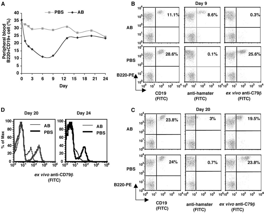
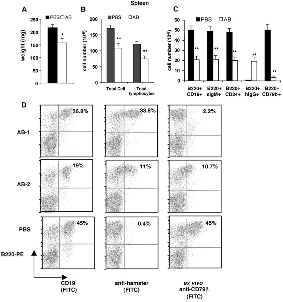
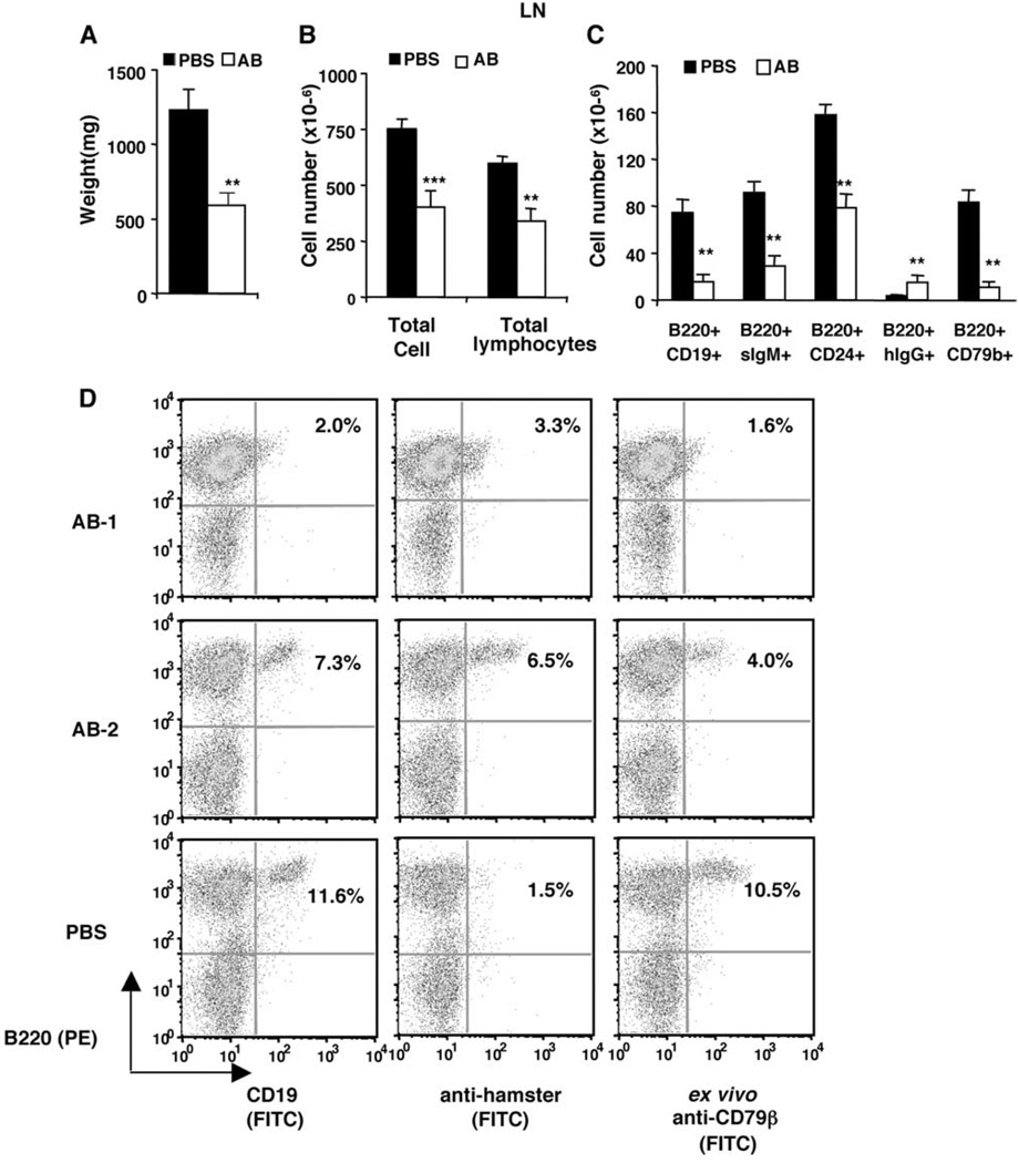
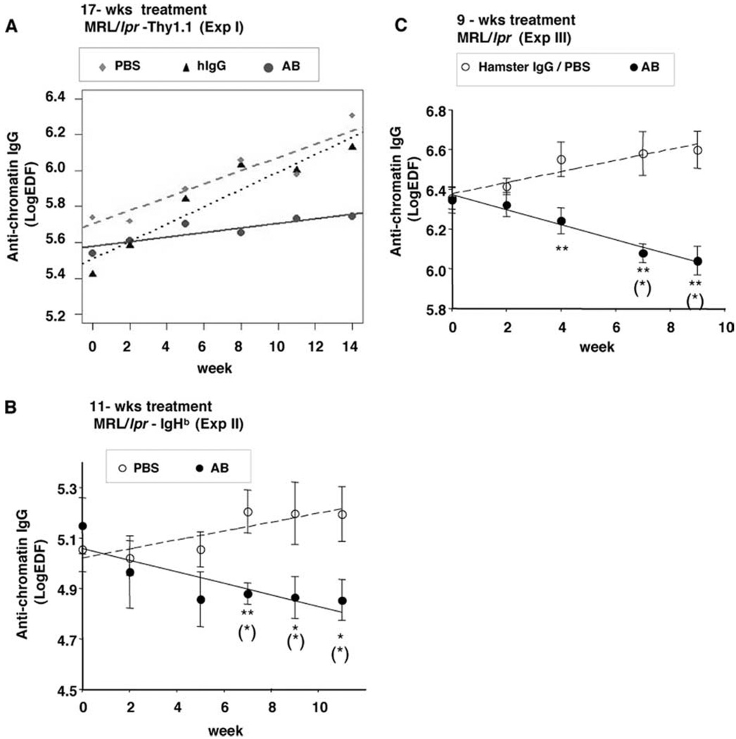
MRL/lpr mice develop a spontaneous systemic lupus erythematosus-like autoimmune syndrome due to a dysfunctional Fas receptor, with contributions from other less well-defined genetic loci. The removal of B cells by genetic manipulation not only prevents autoantibody formation, but it also results in substantially reduced T cell activation and kidney inflammation. To determine whether B cell depletion by administration of Abs is effective in lupus mice with an intact immune system and established disease, we screened several B cell-specific mAbs and found that a combination of anti-CD79alpha and anti-CD79beta Abs was most effective at depleting B cells in vivo. Anti-CD79 therapy started at 4-5 mo of age in MRL/lpr mice significantly decreased B cells (B220(+)CD19(+)) in peripheral blood, bone marrow, and spleens. Treated mice also had a significant increase in the number of both double-negative T cells and naive CD4(+) T cells, and a decreased relative abundance of CD4(+) memory cells. Serum anti-chromatin IgG levels were significantly decreased compared with controls, whereas serum anti-dsDNA IgG, total IgG, or total IgM were unaffected. Overall, survival was improved with lower mean skin scores and significantly fewer focal inflammatory infiltrates in submandibular salivary glands and kidneys. Anti-CD79 mAbs show promise as a potential treatment for systemic lupus erythematosus and as a model for B cell depletion in vivo.


Conflict of interest statement

Disclosures

The authors have no financial conflicts of interest.

Figures

FIGURE 1
FIGURE 1
B cell depletion and recovery in peripheral blood. MRL/lpr-IgHb mice were given one i.p. injection of 0.5 mg each of anti-CD79α and Anti-CD79β (AB) or PBS (n = 2 mice/group). Peripheral blood lymphocytes were collected and analyzed at the indicated time points postinjection. A, Mean of the percentage of peripheral lymphocytes that were B220+CD19+. B and C, Percentage and staining pattern of peripheral blood lymphocytes harvested on day 9 and day 20 that were B220+CD19+, B220+ cells with surface-bound hamster Ab, or B220+ cells that bound ex vivo FITC-Anti-CD79β. D, Expression level of CD79β indicated by fluorescence intensity of ex vivo-bound Anti-CD79β.
FIGURE 2
FIGURE 2
Effects of weekly injection of anti-CD79 mAb on peripheral blood B cells. MRL/lpr-Thy1.1 mice were given weekly i.p. injections of either PBS, control hamster IgG Abs (Hamster IgG), or 0.5 mg each of anti-CD79α and Anti-CD79β (AB) for 17 wk (Table II, Expt. I). Shown are means and SEM of the percentages of peripheral blood lymphocytes that were (A) B220+CD19+ cells; (B) B220+ cells with surface-bound hamster Ab (B220+hIgG+), as detected by anti-hamster Ab; and (C) B220+ cells that bound ex vivo added FITC-anti-CD79β (B220+CD79b+). PBS (n = 3–5 mice); hamster IgG group (n = 3–6); AB (n = 6–8). Numbers of mice were less at later time points either because mice were deceased or sacrificed to examine kidney histology. Except for week 0, the differences between AB and control groups were significant at p < 0.00001 for each time point.
FIGURE 3
FIGURE 3
B cell depletion from spleen. MRL/lpr-IgHb mice were given 11 weekly injections of PBS (n = 7) or 0.5 mg each of anti-CD79α and anti-CD79β (AB, n = 8) (Table II, Expt. II) and were analyzed on week 12 for B cell depletion from lymphoid organs. Shown are means and SEM of (A) spleen weight, (B) total splenic WBC and lymphocyte numbers, and (C) absolute number of B cells. D, Representative anti-hamster IgG staining and binding to ex vivo anti-CD79β by splenocytes from anti-CD79-treated mice (AB-1 and AB-2) or control (PBS). Significant differences (*, p < 0.05; **, p < 0.01) were between AB and control-treated group.
FIGURE 4
FIGURE 4
B cell depletion from lymph nodes. Data from the same experiment as described in Fig. 3. A–C, Means and SEM of weight, lymphocyte numbers, and B cell numbers of lymph nodes. D, Representative anti-hamster IgG staining and binding to ex vivo Anti-CD79β by lymph node cells. Significant differences (*, p < 0.05; **, p < 0.01; ***, p < 0.001) were between AB and control-treated group.
FIGURE 5
FIGURE 5
B cell depletion from bone marrow. Data from the same experiment as described in Fig. 3. A, Percentages of bone marrow B cells. Shown are means and SEM. B, Representative anti-hamster IgG staining and binding to ex vivo Anti-CD79β by bone marrow cells. Significant differences (*, p < 0.05; **, p < 0.01) were between AB and control-treated group.
FIGURE 6
FIGURE 6
B cell subset sensitivity to anti-CD79-mediated depletion correlates with CD79β expression pattern. MRL/lpr-Thy1.1 female were given six weekly i.p. injections of either PBS or 0.5 mg each of anti-CD79α and Anti-CD79β Abs (AB) (Table II, Expt. IV; n = 5 mice/group). At week 7, composition of splenic B cells was analyzed by staining cells with Abs against CD19, CD21, CD23, and CD79β or CD19 and IgD. CD19+ splenic B cells were gated further for follicular (FO), marginal zone (MZ), and newly formed (NF) B cells based on their CD21 and CD23 expression levels. A–C, Percentage and absolute cell number of B cell subsets. Shown are means and SEM. D, Low expression of CD79β in newly formed B cells (CD19+CD21lowCD23low). Shown are representative CD79β staining pattern of splenic FO, MZ, and NF B cells from control. NC (negative control) were splenic lymphocytes incubated with anti-CD19, anti-CD21, and anti-CD23 but not with Anti-CD79β Ab. Significant differences (*, p < 0.05; **, p < 0.01) were between AB and control-treated group.
FIGURE 7
FIGURE 7
Splenic T cell changes in anti-CD79-treated MRL/lpr. Data were from same experiment as described in Fig. 6. A, Relative frequency of T cell subsets. B, Absolute number of T cell subsets. C, Gating of activated, memory, and naive subsets based on CD62L and CD44 expression. Shown are representative data from a pair of mice. D, Relative frequency of CD4+ T cell subset. E, Absolute number of naive CD4+ T cells. Shown are means and SEM. Significant differences (*, p < 0.05; **, p < 0.01) were between AB and control-treated group.
FIGURE 8
FIGURE 8
Anti-CD79 treatment reduced anti-chromatin IgG. Serum anti-chromatin IgG level of MRL/lpr treated for 17 wk (A), 11 wk (B), or 9 wk (C) with PBS, 0.5 mg each of anti-CD79α and Anti-CD79β Abs (AB), or control Abs (Hamster IgG) (Table II, Expts. I, II, and III, respectively). Symbols are group means and SEM of anti-chromatin IgG serum level at each time point. Linear regression lines were fitted to data by mixed-effect models as described in Materials and Methods. *, p < 0.05, **, p < 0.01, significant difference between AB and control groups. (*), p < 0.05, significant difference as compared with week 0.
FIGURE 9
FIGURE 9
Survival rate and skin lesions. MRL/lpr-Thy1.1 mice were given weekly i.p. injections of PBS, control hamster IgG Abs (Hamster IgG), or 0.5 mg each of anti-CD79α and Anti-CD79β (AB) for 17 wk (Table II, Expt. I). A, Survival rate. p < 0.05 by χ2 test starting at week 14. B, Number of mice in each category of skin lesion during treatment. C, Number of mice in each category of skin lesion for those survived from treatment week 0 to week 11 (0, normal; 1, hair loss only; 2, skin inflammation). All mice were free of skin lesion at time of initiation of treatment. *, p < 0.05, significant difference between AB and control-treated groups by Fisher’s exact test.
FIGURE 10
FIGURE 10
Anti-CD79 reduced inflammation in kidney and submandibular salivary glands. MRL/lpr-Thy1.1 female were given six weekly injections of either PBS or anti-CD79α and anti-CD79β Abs (AB) (Table II, Expt. IV; n = 5 mice/group). Shown are representative H&E-stained sections (×100 magnification) of kidneys (A and B) and submandibular salivary glands (C and D). Circled in red are inflammatory infiltrates. b, blood vessel; d, glandular duct. E, Number of focal inflammatory infiltrates in kidneys and submandibular salivary glands. *, p < 0.05 by two-tailed Student’s t test. F, Scores of glomerulonephritis and interstitial nephritis. Significance was determined by two-tailed Fisher’s exact test.

References

    1. Rivera A, Chen CC, Ron N, Dougherty JP, Ron Y. Role of B cells as antigen-presenting cells in vivo revisited: antigen-specific B cells are essential for T cell expansion in lymph nodes and for systemic T cell responses to low antigen concentrations. Int. Immunol. 2001;13:1583–1593. - PubMed
    1. Lipsky PE. Systemic lupus erythematosus: an autoimmune disease of B cell hyperactivity. Nat. Immunol. 2001;2:764–766. - PubMed
    1. Youinou P, Jamin C, Pers JO, Berthou C, Saraux A, Renaudineau Y. B lymphocytes are required for development and treatment of autoimmune diseases. Ann. NY Acad. Sci. 2005;1050:19–33. - PubMed
    1. Lund FE, Garvy BA, Randall TD, Harris DP. Regulatory roles for cytokine-producing B cells in infection and autoimmune disease. Curr. Dir. Autoimmun. 2005;8:25–54. - PubMed
    1. Chan OT, Shlomchik MJ. Cutting edge: B cells promote CD8+ T cell activation in MRL-Faslpr mice independently of MHC class I antigen presentation. J. Immunol. 2000;164:1658–1662. - PubMed

Publication types