. 2008 Sep 1;181(5):3099-107.
doi: 10.4049/jimmunol.181.5.3099.

Radiation-induced CXCL16 release by breast cancer cells attracts effector T cells

Affiliations

Radiation-induced CXCL16 release by breast cancer cells attracts effector T cells

Satoko Matsumura et al. J Immunol. .

Abstract

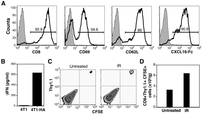
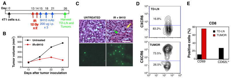
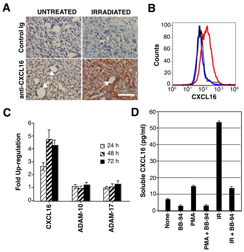
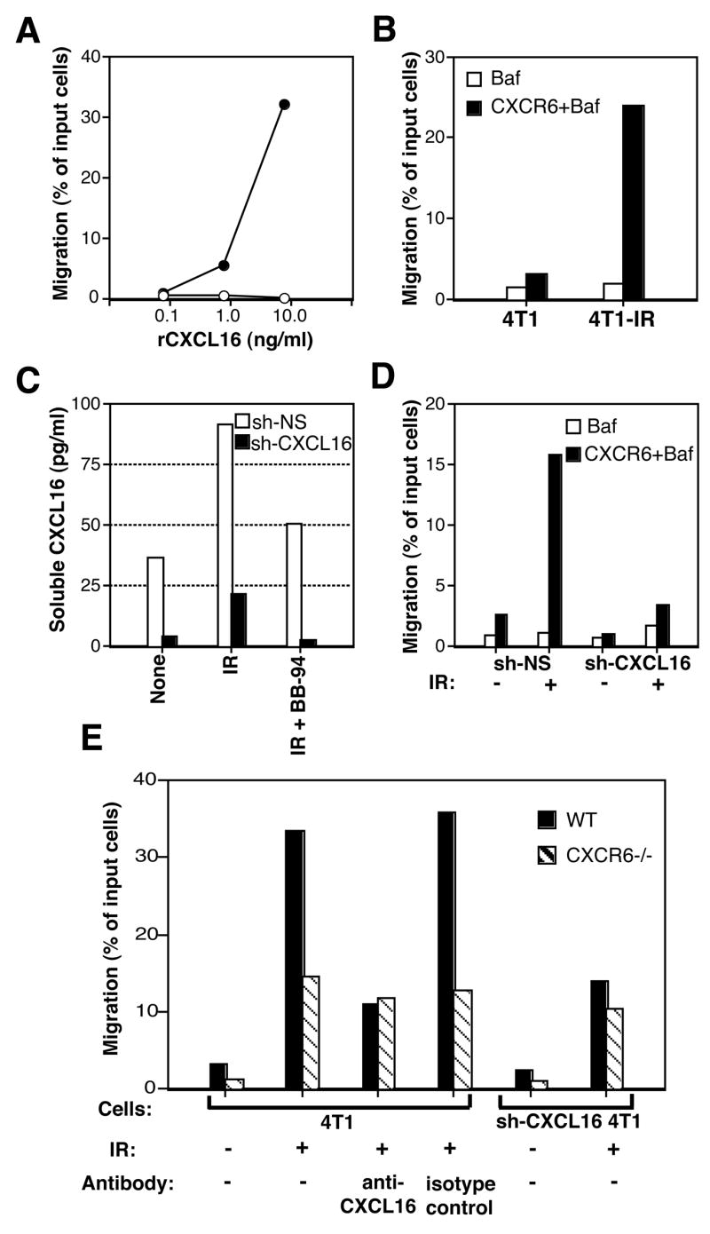
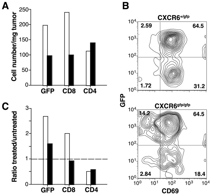
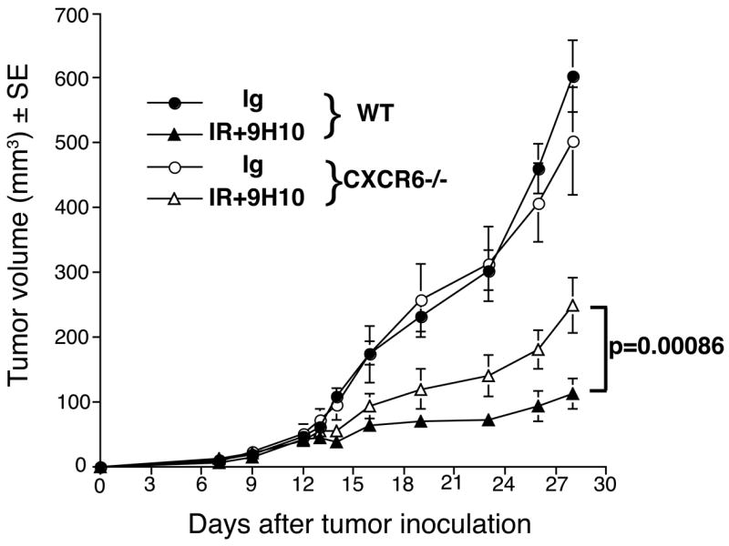
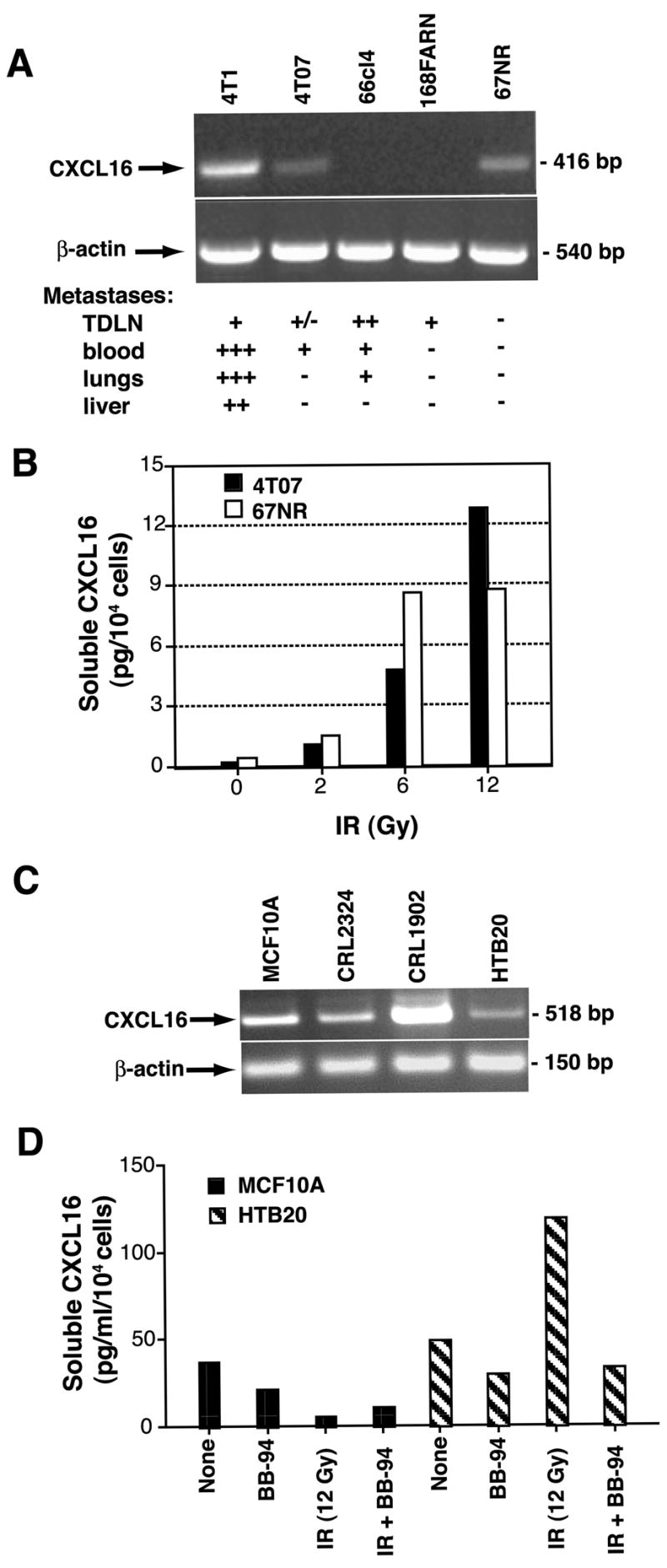
Recruitment of effector T cells to inflamed peripheral tissues is regulated by chemokines and their receptors, but the factors regulating recruitment to tumors remain largely undefined. Ionizing radiation (IR) therapy is a common treatment modality for breast and other cancers. Used as a cytocidal agent for proliferating cancer cells, IR in combination with immunotherapy has been shown to promote immune-mediated tumor destruction in preclinical studies. In this study we demonstrate that IR markedly enhanced the secretion by mouse and human breast cancer cells of CXCL16, a chemokine that binds to CXCR6 on Th1 and activated CD8 effector T cells, and plays an important role in their recruitment to sites of inflammation. Using a poorly immunogenic mouse model of breast cancer, we found that irradiation increased the migration of CD8(+)CXCR6(+) activated T cells to tumors in vitro and in vivo. CXCR6-deficient mice showed reduced infiltration of tumors by activated CD8 T cells and impaired tumor regression following treatment with local IR to the tumor and Abs blocking the negative regulator of T cell activation, CTLA-4. These results provide the first evidence that IR can induce the secretion by cancer cells of proinflammatory chemotactic factors that recruit antitumor effector T cells. The ability of IR to convert tumors into "inflamed" peripheral tissues could be exploited to overcome obstacles at the effector phase of the antitumor immune response and improve the therapeutic efficacy of immunotherapy.


Figures

Figure 1
Figure 1. Irradiation of 4T1 tumors enhances recruitment of adoptively transferred tumor-specific CD8 T cells
WT mice were injected with 4T1-HA cells. Local IR or mock treatment (Untreated) was delivered to the tumors in two 12 Gy fractions at 24 h interval. 48 h after last IR mice received adoptive transfer of CFSE-labeled in vitro activated CL4 T cells. (A) Phenotype of activated CL4 T cells. Shaded histograms are cells stained with control antibody. Numbers indicate the percentage of cells in the gate. (B) Activated CL4 T cells produced IFNγ in response to 4T1-HA ut not parental 4T1 cells. (C, D) Tumors were harvested 24 h after adoptive transfer, and obtained single cell suspensions stained with PE-Cy5-anti-CD8 and PE-anti-Thy1.1 mAbs to identify the transferred T cells. (C) T cells recovered from the tumors did not proliferate within this time as indicated by lack of CFSE dilution. (D) Recruitment of T cells to tumors was enhanced by radiation. Data are from 4 mice of each group. Errors bars are absent because pooling of tumors within each group was necessary to count the CD8 T cells. Results are representative of two experiments.
Figure 2
Figure 2. CD8 TIL in regressing 4T1 tumors from CXCR6+/gfp mice treated with local IR and CTLA-4 blockade express CXCR6
(A) Treatment schedule. (B) Growth of representative tumors from untreated (black squares) and IR+9H10-treated (red diamonds) mice. (C, D) CXCR6+ (as detected by GFP-positivity) TIL in treated tumors. Each tumor was bisected and half was processed for microscopic evaluation whereas the other half was used for flow cytometric analysis. For the latter, tumors from mice in the same group were pooled. (C) Sequential sections of representative tumors from untreated or treated (IR+9H10) mice were stained with H&E (upper panels) or analyzed by fluorescence microscopy for the presence of GFP+ cells (lower panels). In tumors from treated mice residual tumor cells (yellow arrow) were admixed with TIL (green arrow), and many of the TIL were GFP+. Bar, 50 μm. (D, E) Flow cytometric analysis of lymphocytes within dissociated tumors and TD-LN of treated mice. (D) Cells were stained with PE-Cy5-anti-CD8α and gated on CD8+ cells. Numbers indicate the percentage of cells in each gate. (E) Bars show the percentage of CD8+GFP+ cells from tumors (red) and TDLN (black) expressing CD69 or CD62L. Results are from 4 to 8 mice per group, and are representative of 3 experiments.
Figure 3
Figure 3. Release of CXCL16 by 4T1 cells is mediated by a MPase and enhanced by radiation
(A) Up-regulation of CXCL16 by local IR of 4T1 tumors in vivo. Tumors were harvested 48 h after IR (IRRADIATED) or mock treatment (UNTREATED) and stained with anti-CXCL16 or control Ig to detect expression of CXCL16. White arrows indicate blood vessels. Bar, 50 μm. (B) MPase inhibition leads to accumulation of CXCL16 molecules on the surface of 4T1 cells. Cells were incubated for 2 h at 37°C in the presence (red line) or absence (blue line) of BB-94 (20 μM), and stained with anti-CXCL16, or with control Ig (black line), followed by FITC-Donkey anti-Goat Ab. (C) Real time RT-PCR measurement of the expression of CXCL16, ADAM-10 and ADAM-17 in 4T1 cells at different times post-radiation. Samples were normalized to eIF4G II, and expression on untreated cells was assigned a relative value of 1.0. Data are expressed as the mean ±SD (n=3). (D) Soluble CXCL16 was measured by ELISA in supernatants of untreated or irradiated (12 Gy, IR) 4T1 cells cultured for 4 h in the presence or absence of MPase activators/inhibitors, as indicated. Data are expressed as the mean ±SD (n=3).
Figure 4
Figure 4. CXCL16/CXCR6 interactions regulate the migration of CD8 activated T cells to irradiated 4T1 cancer cells in vitro
Results of migration assays are presented as the percentage of input cells migrating to the lower chamber of a transwell filter. (A) Response of Baf-3 (empty circles) and CXCR6+Baf-3 (full circles) cells to various concentrations of rCXCL16. (B) Migration of Baf (white bars) and CXCR6+Baf-3 (black bars) cells towards untreated (4T1) or irradiated with 12 Gy 48 h earlier (4T1-IR) 4T1 cells. (C) 4T1 cells transduced with control (sh-NS, white bars) and silencing (sh-CXCL16, black bars) retroviral vectors were left untreated (None) or irradiated (IR). CXCL16 release was tested in supernatants after 4 h culture in the presence or absence of BB-94. (D) Migration of Baf (white bars) and CXCR6+Baf-3 (black bars) to 4T1 cells transduced with sh-NS and sh-CXCL16. (E) Migration of in vitro activated CD8 T cells from WT (black bars) and CXCR6gfp/gfp (CXCR6−/−, shaded bars) mice to 4T1 and sh-CXCL16-4T1 cells, untreated (−) or irradiated with 12 Gy 48 hours earlier (+). In some wells 1 μg/ml of anti-CXCL16 or isotype control Ab was added, as indicated. Data are representative of two experiments.
Figure 5
Figure 5. CXCR6-deficient mice cannot increase CD8 TIL following treatment with IR and CTLA-4 blockade
CXCR6+/gfp (white bars) and CXCR6gfp/gfp (black bars) mice were injected s.c. with 4T1 cells and left untreated or treated as described in figure 2A. On Day 26 tumors and TDLN were collected and obtained single cell suspensions stained with PE-Cy5-anti-CD3, and PE-anti-CD8 or anti-CD4 mAbs, followed by flow cytometry analysis. The lymphocyte gate was set based on the scattered plots in TDLN. (A) The percentage of cells in the lymphocyte gate positive for CD3, CD3 and CD8 (CD8), CD3 and CD4 (CD4), and GFP was multiplied for the percentage of cells in the lymphocyte gate and for the total number of viable cells isolated from the tumors, and divided for the tumor weight to obtain the number of cells per mg of tumor. (B) TIL isolated from treated mice were gated on CD8+ cells and analyzed for the expression of GFP and CD69, as indicated. Numbers are the percentage of cells in each quadrant. (C) For each marker, the percentage of positive cells in the lymphocyte gate in tumors isolated from treated mice was divided by the percentage of cells isolated from untreated mice. A ratio of one (line) indicates no change, > 1 indicates an increase in treated tumors, and < 1 indicates a decrease in treated tumors. Data are the mean of 8 mice of each genotype per treatment group. Errors bars are absent because pooling of tumors within each group was necessary to count the T cells. Results are representative of two experiments.
Figure 6
Figure 6. Comparison between WT and CXCR6gfp/gfp mice with established 4T1 carcinoma in the response to treatment with local IR and CTLA-4 blockade
Treatment was started on Day 12 post-s.c. inoculation of 4T1 cells in the flank. RT was delivered in two fractions of 12 Gy to the s.c. tumors on Day 12 and 13. Ab were given i.p. 1, 4 and 7 days post-IR. Tumor volume is shown as the mean ± SE in each treatment group up to Day 28 when all animals were alive. Tumor growth was not significantly different (p=0.9) in WT (full circles, n=8) and CXCR6gfp/gfp (empty circles, n=7) mice receiving the control IgG. In contrast, CXCR6gfp/gfp mice receiving IR+9H10 (empty triangles, n=7) had a significantly (p=0.00086) higher tumor volume than WT mice receiving IR+9H10 (full triangles, n=7). Tumor volume differences between RT+9H10 and control (IgG) mice were statistically significant (p<0.0001) within each genotype.
Figure 7
Figure 7. Expression of CXCL16 is common in mouse and human breast cancer lines
(A) CXCL16 was expressed in 3 out of 5 mouse mammary carcinoma lines of differing metastatic potential (26) tested by RT-PCR. (B) IR enhanced in a dose-dependent fashion the release of soluble CXCL16 by the non-metastatic 67NR and the metastatic 4T07 cells. (C) CXCL16 was expressed in 4 human cell lines derived from primary breast specimens of benign breast (MCF 10A) or invasive breast cancer (CRL-2324, CRL-1902, and HTB-20) tested by RT-PCR. (D) Soluble CXCL16 was measured by ELISA in supernatants of untreated or irradiated (12 Gy, IR) MCF 10A and HTB-20 cells cultured for 6 h in the presence or absence of MPase inhibitor BB-94, as indicated. Radiation increased the release of CXCL16 by the HTB-20 tumor cells but not by the epithelial MCF 10A cells derived from benign breast tissue. Data are the mean of duplicate wells and are representative of two experiments.

References

    1. Simpson AJ, Caballero OL, Jungbluth A, Chen YT, Old LJ. Cancer/testis antigens, gametogenesis and cancer. Nat Rev Cancer. 2005;5:615–625. - PubMed
    1. Rosenberg SA, Sherry RM, Morton KE, Scharfman WJ, Yang JC, Topalian SL, Royal RE, Kammula U, Restifo NP, Hughes MS, Schwartzentruber D, Berman DM, Schwarz SL, Ngo LT, Mavroukakis SA, White DE, Steinberg SM. Tumor progression can occur despite the induction of very high levels of self/tumor antigen-specific CD8+ T cells in patients with melanoma. J Immunol. 2005;175:6169–6176. - PubMed
    1. Gajewski TF, Meng Y, Blank C, Brown I, Kacha A, Kline J, Harlin H. Immune resistance orchestrated by the tumor microenvironment. Immunol Rev. 2006;213:131–145. - PubMed
    1. Jain RK. Transport of molecules, particles, and cells in solid tumors. Annu Rev Biomed Eng. 1999;1:241–263. - PubMed
    1. Overwijk WW, Theoret MR, Finkelstein SE, Surman DR, de Jong LA, Vyth-Dreese FA, Dellemijn TA, Antony PA, Spiess PJ, Palmer DC, Heimann DM, Klebanoff CA, Yu Z, Hwang LN, Feigenbaum L, Kruisbeek AM, Rosenberg SA, Restifo NP. Tumor regression and autoimmunity after reversal of a functionally tolerant state of self-reactive CD8+ T cells. J Exp Med. 2003;198:569–580. - PMC - PubMed

Publication types

MeSH terms