. 2008 Sep 1;181(5):3259-67.
doi: 10.4049/jimmunol.181.5.3259.

Colocalization of antigen-specific B and T cells within ectopic lymphoid tissue following immunization with exogenous antigen

Affiliations

Colocalization of antigen-specific B and T cells within ectopic lymphoid tissue following immunization with exogenous antigen

Jason S Weinstein et al. J Immunol. .

Abstract

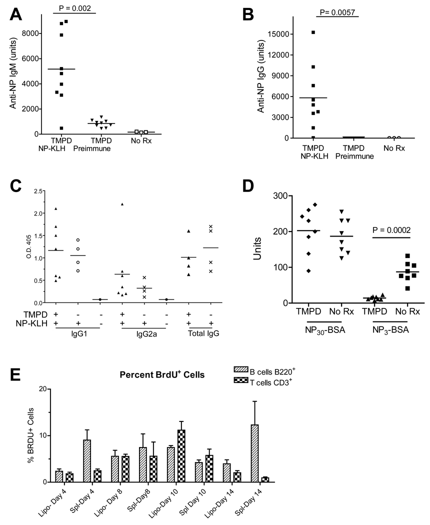
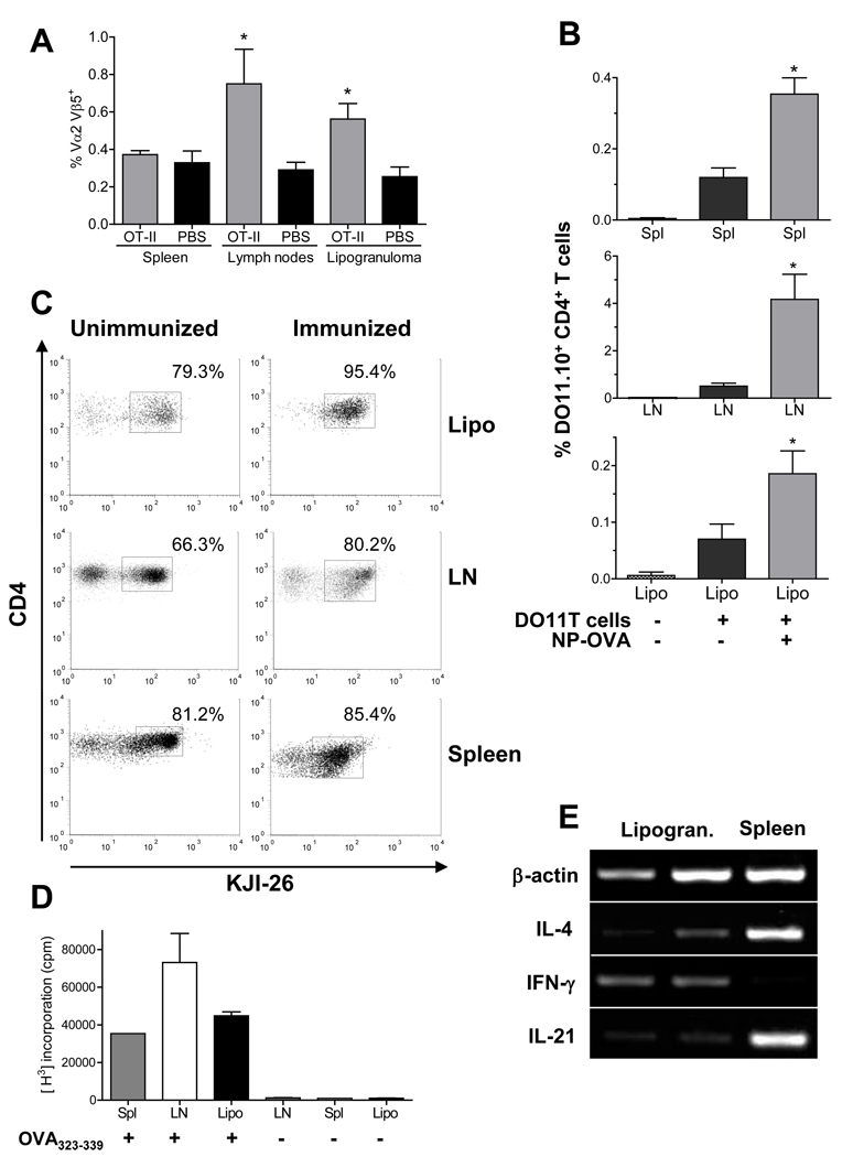
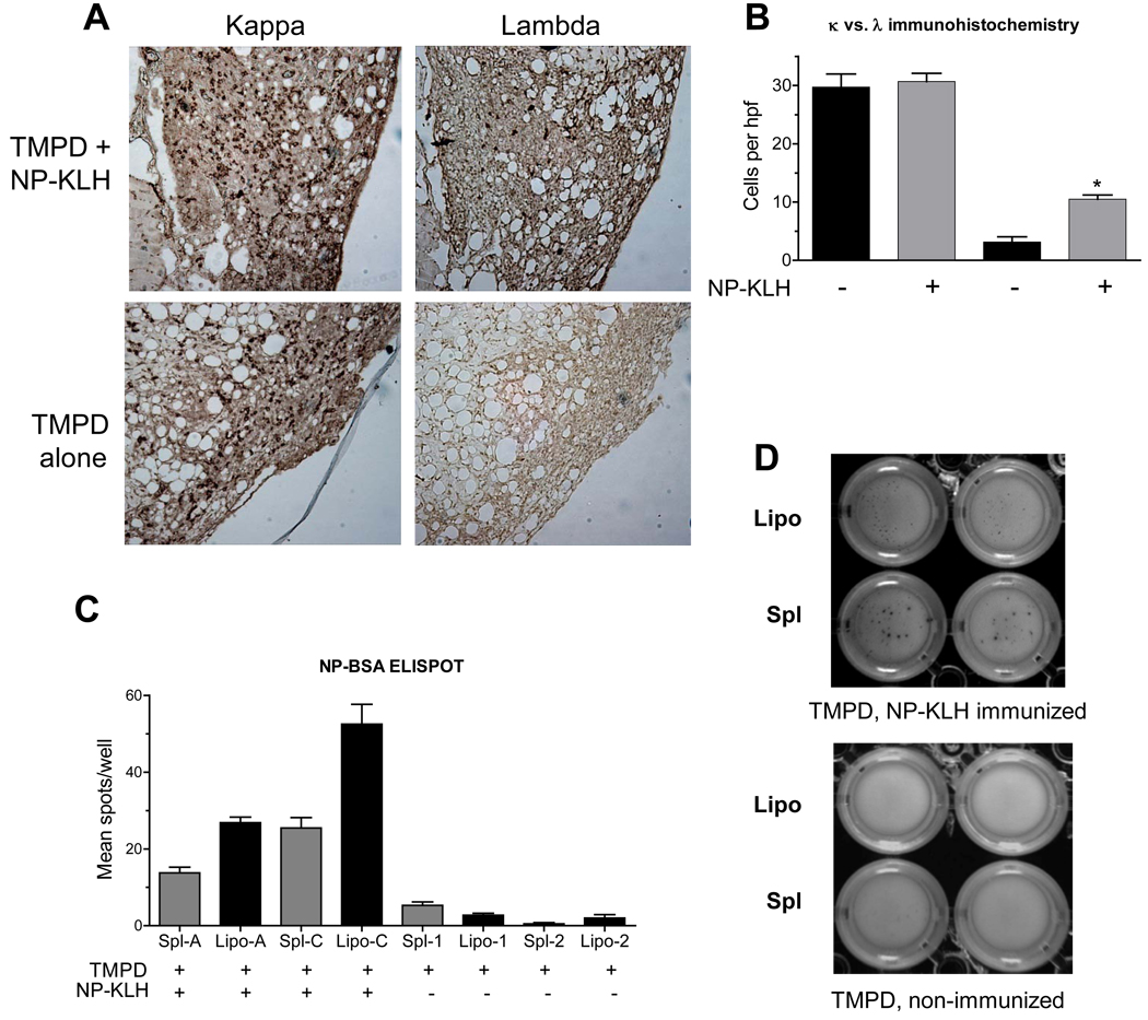
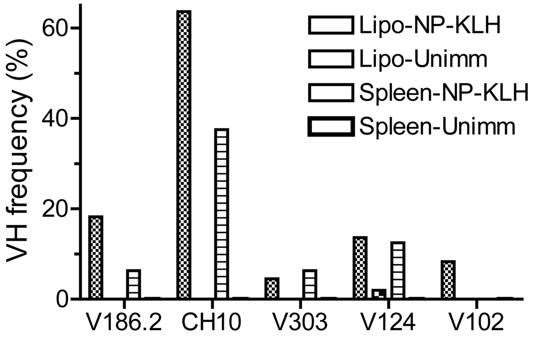
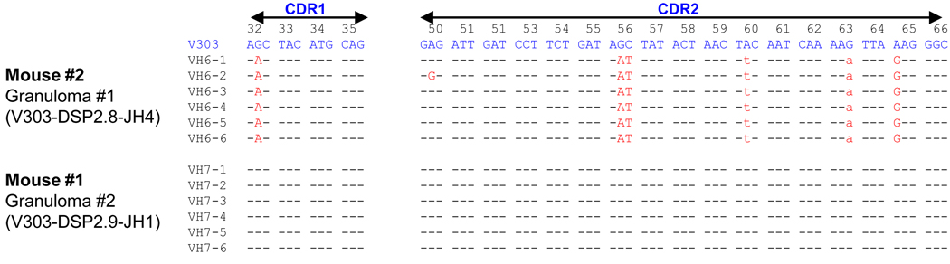
Chronic inflammation promotes the formation of ectopic lymphoid tissue morphologically resembling secondary lymphoid tissues, though it is unclear whether this is a location where Ag-specific immune responses develop or merely a site of lymphocyte accumulation. Ectopic lymphoid tissue formation is associated with many humoral autoimmune diseases, including lupus induced by tetramethylpecadentane in mice. We examined whether an immune response to 4-hydroxy-3-nitrophenyl acetyl-keyhole limpet hemocyanin (NP-KLH) and NP-OVA develops within ectopic lymphoid tissue ("lipogranulomas") induced by tetramethylpecadentane in C57BL/6 mice. Following primary immunization, NP-specific B cells bearing V186.2 and related heavy chains as well as lambda-light chains accumulated within ectopic lymphoid tissue. The number of anti-NP-secreting B cells in the ectopic lymphoid tissue was greatly enhanced by immunization with NP-KLH. Remarkably, the H chain sequences isolated from individual lipogranulomas from these mice were diverse before immunization, whereas individual lipogranulomas from single immunized mice had unique oligo- or monoclonal populations of presumptive NP-specific B cells. H chain CDR sequences bore numerous replacement mutations, consistent with an Ag-driven and T cell-mediated response. In mice adoptively transferred with OT-II or DO11 T cells, there was a striking accumulation of OVA-specific T cells in lipogranulomas after s.c. immunization with NP-OVA. The selective colocalization of proliferating, Ag-specific T and B lymphocytes in lipogranulomas from tetramethylpecadentane-treated mice undergoing primary immunization implicates ectopic lymphoid tissue as a site where Ag-specific humoral immune responses can develop. This has implications for understanding the strong association of humoral autoimmunity with lymphoid neogenesis, which may be associated with deficient censoring of autoreactive cells.


Figures

Figure 1
Figure 1. Serum anti-NP response after immunization with NP-KLH
A and B, B6 mice injected with TMPD 3 months earlier were immunized with NP-KLH. At day 12, sera were tested for IgM (A) and IgG (B) anti-NP antibodies by ELISA using NP-BSA. There was a significant increase in both IgM and IgG anti-NP from pre-immune sera to day 12 immune sera. C, Isotypes of anti-NP antibodies (ELISA) from mice either pre-treated or not pre-treated with TMPD prior to immunization with NP-KLH. D, Affinity of anti-NP antibodies developing in TMPD-treated mice vs. controls (no Rx) immunized with NP-KLH. Serum binding activity (measured in arbitrary units using a standard curve) was measured using NP30-BSA (low and high avidity/affinity antibodies) and NP3-BSA (high avidity/affinity antibodies). E, In vivo BrdU labeling of T and B cells in the lipogranulomas and spleens of TMPD-treated and NP-KLH immunized mice (n = 3). Single cell suspensions were stained with anti-CD45R (B220) and anti-CD3 antibodies and with an anti-BrdU antibody. Data are expressed as the % of BrdU+ B cells or T cells, respectively, at 4, 8, 10, or 14 days after immunization with NP-KLH.
Figure 2
Figure 2. OVA-specific T cells in lipogranulomas
A, Single cell suspensions from spleen, lymph nodes, or lipogranulomas of mice injected i.v. with 5 × 10⁶ OT-II T cells (n = 6) or PBS (n = 5) were analyzed by flow cytometry 7 days after immunization with NP-OVA. The Vα2+Vβ5+ values represent the total percentage of CD3+CD4+ T cells. (* Lymph nodes, P = 0.009; Lipogranuloma, P = 0.02, Mann Whitney test) B, Mice were injected i.v. with DO11.10 T cells (n = 12) or PBS (n = 5) were analyzed by flow cytometry. Some mice that received DO11.10 T cells were immunized with NP-OVA (n = 7) and others were not (n = 5). (* Spleen, P = 0.0006; Lymph node, P = 0.01, Lipogranuloma, P = 0.02, Mann Whitney test) C, Single cell suspensions of lipogranulomas, spleen and draining lymph nodes from immunized or non-immunized mice were gated on live CD3+CD4+ cells (cytox blue) and then the % of KJI-26+ (DO11.10 T cells) was determined from CD3+ CD4+ cells (representative of three independent experiments). D, Isolated CD4+ T cells were cultured with or without OVA323–339 for 72 h. T cell proliferation was measured by [3H] incorporation (representative of three independent experiments). E, cDNA was synthesized from two lipogranulomas (Lipo) or spleen from a TMPD treated mice immunized with NP-KLH. IL-4, IFNγ, and IL-21 expression was determined by RT-PCR and normalized to β-actin expression (representative of four different mice).
Figure 3
Figure 3. Anti-NP B cells in ectopic lymphoid tissue
A, Light chain staining of B cells in lipogranulomas from B6 mice treated with TMPD alone or treated with TMPD and then immunized with NP-KLH. Paraformaldehyde-fixed tissue was analyzed 12 days after NP-KLH immunization. Paraffin sections were stained with anti-κ and anti-λ light chain antibodies. B, Number of κ and λ positive cells per high power field in mice treated with TMPD + NP-KLH immunization or with TMPD alone (* P = 0.02, Mann Whitney test; representative of five independent experiments). C, ELISPOT assay for anti-NP B cells. MultiScreen HTS IP plates containing a 0.45 µm Immobilon-P membrane were coated with 1 µg/mL NP-BSA. Lipogranuloma and spleen cells from TMPD treated mice (n = 2) or TMPD-treated and NP-KLH immunized mice (n = 2) were added to triplicate wells for 24 h before adding biotinylated goat anti-mouse IgM antibodies, streptavidin-peroxidase, and BCIP-NBT substrate. Number of spots per well was determined (* P = 0.03 for both mouse A and mouse B, Mann Whitney test; representative of three independent experiments). D, Relative sizes of individual spots in ELISPOT assays using lipogranuloma (Lipo) or spleen (Spl) cells from TMPD treated mice either with or without NP-KLH immunization.
Figure 4
Figure 4. VH segment usage in lipogranulomas and spleen
Lipogranulomas from a TMPD-treated mouse immunized with NP-KLH were pooled at day 12 and RNA was isolated. Immunoglobulin VH sequences were determined from lipogranulomas (n = 50) and spleen (n = 50) from the same mouse. All of the sequences recovered from lipogranulomas bore V186.2, CH10, V303, V102, or V124 vs. 62.6% of the sequences recovered from the spleen (representative of three independent experiments).
Figure 5
Figure 5. Oligoclonal VH sequences from lipogranulomas of immunized mice
Heavy chain sequences are shown from spleen and lipogranulomas of two mice immunized with NP-KLH (day 12) and two pre-immune (not immunized) mice. V-D-J sequences were amplified from cDNA by PCR and sequenced to determine VH, D, and JH usage. Boxed sequences utilize VH sequences associated with anti-NP reactivity. Related sequences are shown in the same format.
Figure 6
Figure 6. CDR1 and CDR2 sequences from H-chains isolated from lipogranulomas
Sequences of H-chains from Mouse #1, lipogranuloma 2 (V303-DSP2.9-JH1) and Mouse #2, Lipogranuloma 1 (V303-DSP2.8-JH4) are aligned. Somatic mutations are indicated. Replacement mutations capitalized, silent mutations lower case.

References

    1. Kratz A, Campos-Neto A, Hanson MS, Ruddle NH. Chronic inflammation caused by lymphotoxin is lymphoid neogenesis. J.Exp.Med. 1996;183:1461–1472. - PMC - PubMed
    1. Drayton DL, Liao S, Mounzer RH, Ruddle NH. Lymphoid organ development: from ontogeny to neogenesis. Nat.Immunol. 2006;7:344–353. - PubMed
    1. Hjelmstrom P. Lymphoid neogenesis: de novo formation of lymphoid tissue in chronic inflammation through expression of homing chemokines. J.Leukoc.Biol. 2001;69:331–339. - PubMed
    1. Cassese G, Lindenau S, de Boer B, Arce S, Hauser A, Riemekasten G, Berek C, Hiepe F, Krenn V, Radbruch A, Manz RA. Inflamed kidneys of NZB / W mice are a major site for the homeostasis of plasma cells. Eur.J.Immunol. 2001;31:2726–2732. - PubMed
    1. Satoh M, Reeves WH. Induction of lupus-associated autoantibodies in BALB/c mice by intraperitoneal injection of pristane. J.Exp.Med. 1994;180:2341–2346. - PMC - PubMed

Publication types