. 2008 Sep 1;181(5):3495-502.
doi: 10.4049/jimmunol.181.5.3495.

Lipopolysaccharide is a direct agonist for platelet RNA splicing

Affiliations

Lipopolysaccharide is a direct agonist for platelet RNA splicing

Pavel N Shashkin et al. J Immunol. .

Abstract

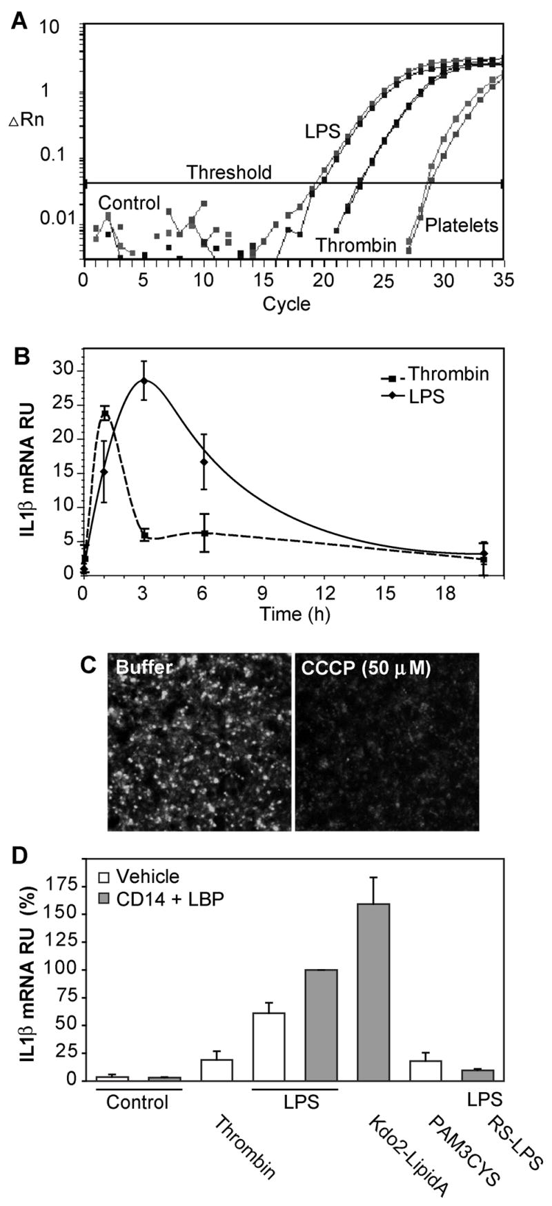
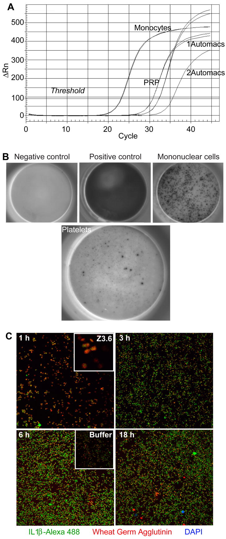
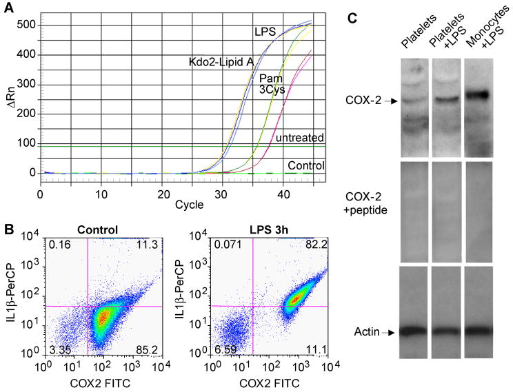
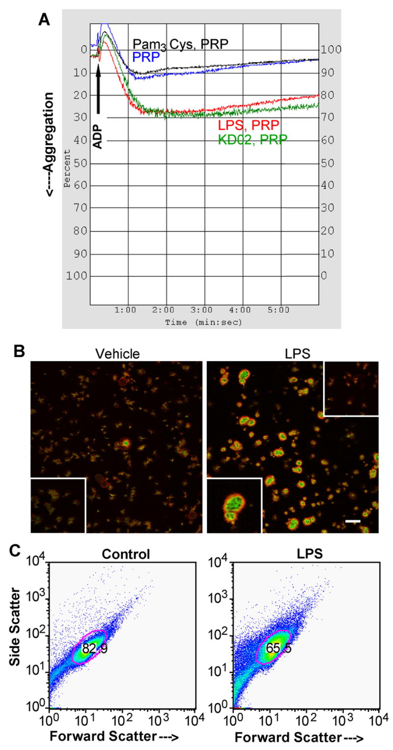
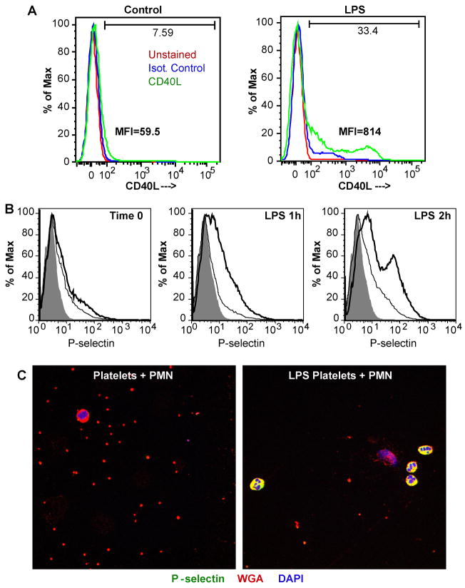
Platelets express TLR4 receptors, but its ligand LPS does not directly activate thrombotic functions nor, obviously, transcription by these anucleate cells. Platelets, however, store information that changes their phenotype over a few hours in the form of unprocessed RNA transcripts. We show even low concentrations of LPS in the presence of soluble CD14 initiated splicing of unprocessed IL-1beta RNA, with translation and accumulation of IL-1beta protein. LPS was a more robust agonist for this response than thrombin. Platelets also contained cyclooxygenase-2 pre-mRNA, which also was spliced and translated after LPS stimulation. Flow cytometry and immunocytochemistry of platelets extensively purified by negative immunodepletion showed platelets contained IL-1beta, and quantitative assessment of white blood cell contamination by CD14 real time PCR confirms that leukocytes were not the IL-1beta source, nor were they required for platelet stimulation. LPS did not initiate rapid platelet responses, but over time did prime platelet aggregation to soluble agonists, induced actin rearrangement, and initiated granule secretion with P-selectin expression that resulted the coating of quiescent leukocytes with activated platelets. LPS is a direct agonist for platelets that allows these cells to directly participate in the innate immune response to bacteria.


Figures

Figure 1
Figure 1. LPS induces IL-1β RNA processing in purified platelets
A. Real-time RT-PCR progression curves for spliced IL-1β RNA. Logarithmic accumulation of IL-1β amplicons using primers anchored in exon 1 and 3. Duplicate determinations using RNA extracted from platelets (8 ng total RNA) left untreated, or stimulated with 0.1 U thrombin for 30 min, or 100 ng/ml of LPS along with 150 ng/ml of recombinant CD14 and 100 ng/ml LPS-binding protein (LBP) for 3 hours. ▵Rn is the magnitude of the signal normalized to total RNA. B. Kinetics of IL-1β mRNA accumulation. The accumulation of spliced IL-1β RNA in purified human platelets treated with 0.1 u/ml thrombin or LPS in the presence of soluble CD14 and LBP (in three separate experiments ± SE) compared to the basal amount by quantitative RT-PCR as in the above panel. C. Platelets remain viable after prolonged stimulation. Accumulation of the cationic dye MitoTracker Red®, with or without subsequent dissipation of the mitochondrial transmembrane potential with CCCP, was imaged after 20 h of LPS exposure. D. LPS stimulation of platelet RNA splicing requires TLR4 receptor components. Purified platelets were stimulated with 100 ng/ml LPS, 100 ng/ml KDO2-Lipid A, 100 μg/ml Pam3Cys-SK4, or 0.1 units of thrombin before RNA isolated 3 hours later. Some samples contained soluble CD14 and LBP, while the competitive TLR4 receptor antagonist Rhodobacter sphaeroides LPS (RS-LPS) was present in one sample as stated. Bars represent standard errors. N = 4.
Figure 2
Figure 2. LPS treatment induces IL1β expression
A. Platelet IL-1β accumulation is time dependent. Purified platelets were treated with 100 ng/ml of E. coli LPS, recombinant CD14 and recombinant LBP for the stated times as in Fig. 1, permeabilized and stained with either anti-IL-1β or an isotype matched control antibody and then analyzed by flow cytometry as described in “Materials and Methods”. Both non-immune and anti-IL-1β antibodies were used at time 0 to define background binding, the shaded area and the light gray trace, that were equivalent. The ordinate is the number of events normalized to the maximal number of counts, the abscissa is the fluorescent channel 1 bin. B. LPS concentration/response profile. Platelets were treated with the stated concentration of LPS, along with recombinant CD14 and LBP, for 1 hour, and then assessed for IL-1β expression as above. Representative of four independent experiments.
Figure 3
Figure 3. Platelets are a direct target of LPS stimulation
A. Quantitative assessment of the effectiveness of platelet purification by immunodepletion of leukocytes. Platelet rich plasma (PRP) was purified using anti-CD14, -CD15, -CD45 and anti-glycophorin coated magnetic beads by passage once or twice over a Miltenyi AutoMACS column. RNA was extracted from the recovered cells and normalized for total RNA content before the content of CD14 RNA was quantitatively assessed in duplicate samples by real-time PCR as described in “Materials and methods”. B. ELISpot detection of IL-1β producing cells in a preparation of purified platelets. The top leftmost panel is a negative buffer control, 0.8 ng recombinant IL-1β (top middle), and 20,000 density purified monocytes stimulated by LPS (top right). The right panel shows 107 platelets stimulated by 100 ng LPS for 17 hours. C. Confocal microscopy of cells in a platelet preparation expressing IL-1β. Platelets suspended over gelatin-coated cover slips were treated with 100 ng/ml LPS for the stated times, permeabilized, and then stained with mouse anti-IL-1β and Alexa 488-conjugated anti-mouse antibody (green) and Alexa 594-conjugated wheat germ agglutinin (WGA) that stains plasma membrane sialated proteins and gangliosides (red). The fixative contained the nuclear stain DAPI to identify contaminating white blood cells (blue). Upper inset: Higher magnification (3.7×) shows IL-1β is localized as punctate intracytoplasmic inclusions at times shortly after LPS addition. Lower inset: 24 h of incubation in the absence of LPS reveals lower levels of IL-1β staining. Representative of three experiments.
Figure 4
Figure 4. Platelets express cyclooxygenase-2 after LPS stimulation
A. Progression curve of real-time PCR for spliced cyclooxygenase-2 RNA. Purified platelets were stimulated with 100 ng E. coli LPS, 100 ng KDO2-Lipid A, 100 μg Pam3Cys-SK4 or left untreated. RNA was extracted and tested as in Figure 3 using primers anchored exon 3 and the junction formed by splicing exons 3 and 4. B. Platelet expression of IL-1β and cyclooxygenase-2. Purified platelets were stimulated with 100 ng/ml E. coli LPS for 3h or left untreated, permeabilized, fixed and stained with a combination of mouse anti-IL-1β and PerCP-conjugated goat anti-mouse antibody followed by a directly conjugated FITC-anti-cyclooxygenase-2 antibody. The cells were subjected to flow cytometry, gated by forward and side scatter, and FITC and PerCP fluorescence was assessed in channels FL1 and FL3, respectively. This experiment has been replicated more than fifteen times, although the level of cyclooxygenase-2 expression varied among donors. C. Cyclooxygenase-2 immunoblot in extracts of purified platelets. Platelets or density-purified monocytes were treated with LPS, recombinant soluble CD14 and LPS binding protein as in the preceding panel for 17 h, while monocytes were treated with 100 ng/ml LPS alone for 17 h. Upper panel, anti-cyclooxygenase-2 western blot. Middle panel, anti-cyclooxygenase-2 western blot using antibody pre-incubated with its cognate peptide. Lower panel, β-actin western blot.
Figure 5
Figure 5. LPS primes platelet aggregation and alters platelet structure
A. LPS primes ADP-induced aggregation in platelet-rich plasma (PRP). LPS (100 ng), KDO2-Lipid A (100 ng) or Pam3Cys-SK4 (100 μg) was added to 1 ml of PRP and incubated for 1h before addition to a stirred aggregometer cuvette. As shown by the arrow, 20 μM ADP was added and light transmittance was assessed over the subsequent 6 min. LPS did not induce aggregation without the addition of ADP (not shown). B. Polymerized actin staining of adherent platelets. Confocal microscopy images of control (left) and platelets stimulated with 100 ng/ml E. coli LPS (right) in the presence of autologous serum or soluble CD14 and LPS binding protein for 3h. Green staining is polymerized (F-actin) stained by FITC-phalloidin and red fluorescence is unpolymerized G-actin. The bar is 20 μm. Lower inserts: 2× magnified fields of respective images. Upper insert: pretreatment with cytochalasin B abrogated actin polymerization. C. LPS affects platelet structure. Platelets were treated or not with LPS for 3 h as in the preceding panel and then analyzed by flow cytometry for forward scatter (size) and side scatter (granularity). The area of the gate imposed on the dot blot stained for the platelet protein CD42b. This experiment has been repeated more than twenty times.
Figure 6
Figure 6. LPS induces degranulation and P-selectin expression that promotes neutrophil binding
A. CD40L surface expression. Platelets were stimulated by LPS, or not, for 3h, fixed and stained with either mouse anti-CD40L or an isotype matched control antibody before the cells were analyzed by flow cytometry. MFI, mean fluorescence intensity. B. P-selectin surface expression. Platelets were treated with buffer or 100 ng/ml LPS for 1 or 2 hours before fixation, antibody staining, and flow cytometry as before. The shadowed area shows unstained platelets, the thin line defines isotype control antibody staining, while the bold line shows P-selectin (CD62P) staining. C. LPS-stimulated platelets decorate quiescent neutrophils. Platelets were treated with buffer or LPS for 2.5 h as before, washed, and then suspended with quiescent autologous neutrophils for 10 min on gelatin-coated slides, fixed, and stained for P-selectin (green FITC fluorescence), plasma membrane by Alexa 594-conjugated wheat germ agglutinin (red), and nuclei with DAPI (blue). The images are representative of three experiments.

References

    1. Palsson-McDermott EM, O’Neill LA. Signal transduction by the lipopolysaccharide receptor, Toll-like receptor-4. Immunology. 2004;113:153–162. - PMC - PubMed
    1. Andonegui G, Kerfoot SM, McNagny K, Ebbert KV, Patel KD, Kubes P. Platelets express functional Toll-like receptor-4. Blood. 2005;106:2417–2423. - PubMed
    1. Cognasse F, Hamzeh H, Chavarin P, Acquart S, Genin C, Garraud O. Evidence of Toll-like receptor molecules on human platelets. Immunol Cell Biol. 2005;83:196–198. - PubMed
    1. Aslam R, Speck ER, Kim M, Crow AR, Bang KW, Nestel FP, Ni H, Lazarus AH, Freedman J, Semple JW. Platelet Toll-like receptor expression modulates lipopolysaccharide-induced thrombocytopenia and tumor necrosis factor-alpha production in vivo. Blood. 2006;107:637–641. - PubMed
    1. Stahl AL, Svensson M, Morgelin M, Svanborg C, Tarr PI, Mooney JC, Watkins SL, Johnson R, Karpman D. Lipopolysaccharide from enterohemorrhagic Escherichia coli binds to platelets through TLR4 and CD62 and is detected on circulating platelets in patients with hemolytic uremic syndrome. Blood. 2006;108:167–176. - PMC - PubMed

Publication types