. 2008 Oct;16(10):1710-8.
doi: 10.1038/mt.2008.166. Epub 2008 Aug 19.

Expanded repertoire of AAV vector serotypes mediate unique patterns of transduction in mouse brain

Affiliations

Expanded repertoire of AAV vector serotypes mediate unique patterns of transduction in mouse brain

Cassia N Cearley et al. Mol Ther. 2008 Oct.

Abstract

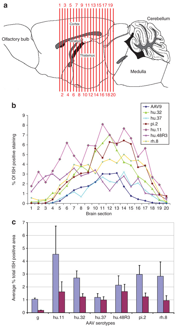
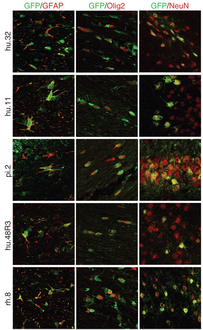
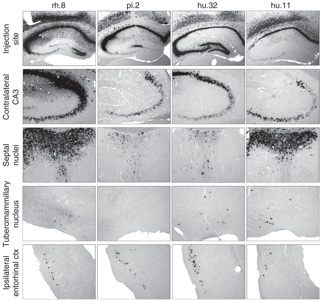
A wide diversity of adeno-associated virus (AAV) structural proteins uncovered from latent genomes in primate tissue has expanded the number of AAV vector serotypes, which can potentially confer unique cell tropism to the vector. We evaluated 17 of these vectors in the mouse brain using green fluorescent protein (GFP) as a reporter gene. A rapid initial evaluation was performed by neonatal lateral ventricle injections. Vectors made with capsids hu.32, hu.37, pi.2, hu.11, rh.8, hu.48R3, and AAV9 for comparison were selected for further analysis based on their ability to transduce large numbers of cells and result in novel patterns of cell transduction. These vectors were injected into adult brains in four major structures (cortex, striatum, hippocampus, and thalamus), and all were found to transduce neurons. In addition, hu.32, hu.11, pi.2, hu.48R3, and rh.8 resulted in GFP expression in some astrocytes or oligodendrocytes. AAVs rh.8, pi.2, hu.32, and hu.11 also appeared to result in neuronal transport of the vector genome. Vector transport was studied by a single unilateral injection into the hippocampus and vector genome was found in projection sites of the hippocampus. These unique patterns of cell transduction expand the potential repertoire for targeting AAV vectors to selected subsets of brain cells.


Figures

Figure 1
Figure 1. Phylogenetic tree of all tested adeno-associated virus (AAV) isolates
Dendrogram, using the Neighbor-Joining method and Poisson correction, was made with molecular evolutionary genetics analysis software (The Biodesign Institute, Tempe, AZ). AAVs are grouped in their respective clades as noted by brackets and the closest known serotype relative is shown in blue. Branch lengths correspond to the number of estimated amino-acid substitutions for that distance and the distance scale of branch length is noted on the bottom left.
Figure 2
Figure 2. Comparison of vector distribution resulting from injections of adeno-associated virus (AAV) vectors into the lateral ventricles of neonatal mice
Animals (n = 2–3 per vector) were injected with 1 µl of AAV vector into each lateral ventricle and killed 2 weeks after injection. The highest obtainable vector titers were used for all injections (Table 1). Frozen sections underwent in situ hybridization using a riboprobe against the green fluorescent protein sequence. Images shown are single-hemisphere representative examples of the transduction characteristics of the individual capsid isolates, grouped by clade. Vectors rh.32.33 and rh.8 do not fall into known clades, and hence they are classified as “Other.”
Figure 3
Figure 3. Comparison of vector distribution resulting from injections of novel adeno-associated virus (AAV) vectors into adult mice
AAV9, hu.32, hu.37, pi.2, hu.11, hu.48R3, and rh.8 were injected into the cortex, striatum, hippocampus, and thalamus (1 µ l per injection site) of adult mice (>2 months of age), and animals were killed 3 weeks after injection. The highest obtainable vector titers were used for all injections (Table 1). Frozen sections underwent in situ hybridization (ISH) using a riboprobe against the green fluorescent protein sequence. (a) Representative examples of the transduction capabilities of the individual vectors. The injection locations are shown with arrows on the AAV9 sections. The rostral-caudal level of the coronal section relative to bregma is given with (+) indicating that the location is rostral to bregma and a (−) indicating the section is caudal to bregma. Boxes in a, not drawn to scale, correspond to the panels in b. (b) ISH-positive cells can be found in the contralateral hippocampus of AAV9, hu.32, pi.2, hu.11, and rh.8 injected animals, indicating that these vectors were capable of axonal transport from the injected hippocampus.
Figure 4
Figure 4. Comparison of transduced area after adeno-associated virus (AAV) vector injections into adult animals
The percentage of in situ hybridization (ISH) positive–stained area within 20 equally spaced sections (~240 µm apart) across the transduced region of the injected mouse brains was calculated using a custom-designed Image-Pro macro. (a) Locations of the 20 sections analyzed are shown, the numbers in a indicate the position of the section and correspond with numbers given along the x-axis in b. Drawing after Paxinos and Franklin. (b) The percentage of ISH-stained area for each section was calculated and the numbers were graphed across all 20 sections. (c) The average percent of ISH staining to total area of sections were averaged for all 20 sections and averaged across the three mice per group (blue bars are actual percentages, red bars are percentages normalized to 1 × 10¹³ genome equivalents/ml).
Figure 5
Figure 5. Novel adeno-associated virus (AAV) vectors result in green fluorescent protein (GFP) expression in non-neuronal cells
GFP-positive sections from brains injected with AAV hu.32, hu.11, pi.2, hu.48R3, and rh.8 were co-labeled with antibodies to the astrocytic marker glial fibrillary acidic protein (GFAP), the oligodendrocytic marker Olig2, or the neuronal marker NeuN. The GFP-positive cells stain green, whereas the cell-specific markers fluoresce red via conjugated secondary antibodies. All pictures were taken using confocal microscopy. GFAP and Olig2 pictures were taken with ×63 magnification and a ×2.3 zoom. NeuN pictures were taken with ×40 magnification and ×2 zoom. Pictures showing co-localization with GFAP or Olig2 were taken of the corpus callosum or external capsule. Pictures showing co-localization with NeuN were taken on the outer edges of the transduced regions and are of the hippocampus (AAV9 or pi.2), the thalamus (hu.32, hu.11, or hu.48R3), or of the cortex (rh.8).
Figure 6
Figure 6. Adeno-associated virus (AAV) vector transport following unilateral injection into the hippocampus
The highest obtainable vector titers were used for the injections (Table 1). Animals (n = 3/group) were injected into the unilateral hippocampus with rh.8, pi.2, hu.32, or hu.11, and examined 3 weeks postinjection for vector distribution. Frozen sections underwent in situ hybridization (ISH) using a riboprobe against the green fluorescent protein sequence. The location of the injection is shown for the vectors in the first row of panels. ISH-positive cells could be found in projection sites of the hippocampus including the CA3 region of the contralateral hippocampus, the lateral septal nuclei, the entorhinal cortex, and the tuberomammillary nucleus.

References

    1. Davidson BL, Stein CS, Heth JA, Martins I, Kotin RM, Derksen TA, et al. Recombinant adeno-associated virus type 2, 4, and 5 vectors: transduction of variant cell types and regions in the mammalian central nervous system. Proc Natl Acad Sci USA. 2000;97:3428–3432. - PMC - PubMed
    1. Passini MA, Watson DJ, Vite CH, Landsburg DJ, Feigenbaum AL, Wolfe JH. Intraventricular brain injection of adeno-associated virus type 1 (AAV1) in neonatal mice results in complementary patterns of neuronal transduction to AAV2 and total long-term correction of storage lesions in the brains of beta-glucuronidase-deficient mice. J Virol. 2003;77:7034–7040. - PMC - PubMed
    1. Burger C, Gorbatyuk OS, Velardo MJ, Peden CS, Williams P, Zolotukhin S, et al. Recombinant AAV viral vectors pseudotyped with viral capsids from serotypes 1, 2, and 5 display differential efficiency and cell tropism after delivery to different regions of the central nervous system. Mol Ther. 2004;10:302–317. - PubMed
    1. Cearley CN, Wolfe JH. Transduction characteristics of adeno-associated virus vectors expressing cap serotypes 7, 8, 9, and Rh10 in the mouse brain. Mol Ther. 2006;13:528–537. - PubMed
    1. Taymans JM, Vandenberghe LH, Haute CV, Thiry I, Deroose CM, Mortelmans L, et al. Comparative analysis of adeno-associated viral vector serotypes 1, 2, 5, 7, and 8 in mouse brain. Hum Gene Ther. 2007;18:195–206. - PubMed

Publication types

Substances