. 2008 Aug 20;3(8):e3000.
doi: 10.1371/journal.pone.0003000.

The cellular prion protein PrP(c) is involved in the proliferation of epithelial cells and in the distribution of junction-associated proteins

Affiliations

The cellular prion protein PrP(c) is involved in the proliferation of epithelial cells and in the distribution of junction-associated proteins

Etienne Morel et al. PLoS One. .

Erratum in

  • PLoS ONE. 2008;3(9). doi: 10.1371/annotation/0b364095-9f93-4cb9-9a2e-aae5ed1bf362
  • PLoS ONE. 2008;3(9). doi: 10.1371/annotation/e5a42567-afa3-422a-886e-44ca642c6fe2. Thievend, Cathy Pichol [corrected to Pichol Thievend, Cathy]

Abstract

Background: The physiological function of the ubiquitous cellular prion protein, PrP(c), is still under debate. It was essentially studied in nervous system, but poorly investigated in epithelial cells. We previously reported that PrP(c) is targeted to cell-cell junctions of polarized epithelial cells, where it interacts with c-Src.

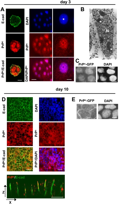
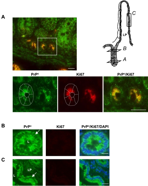
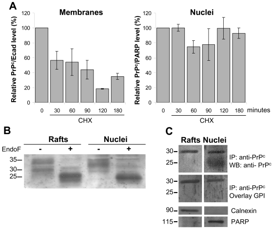
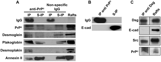
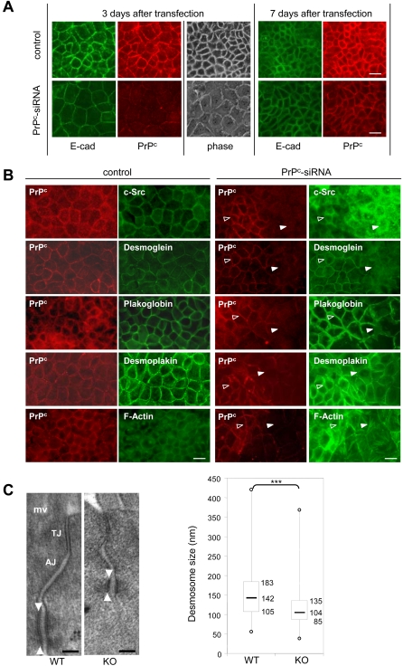
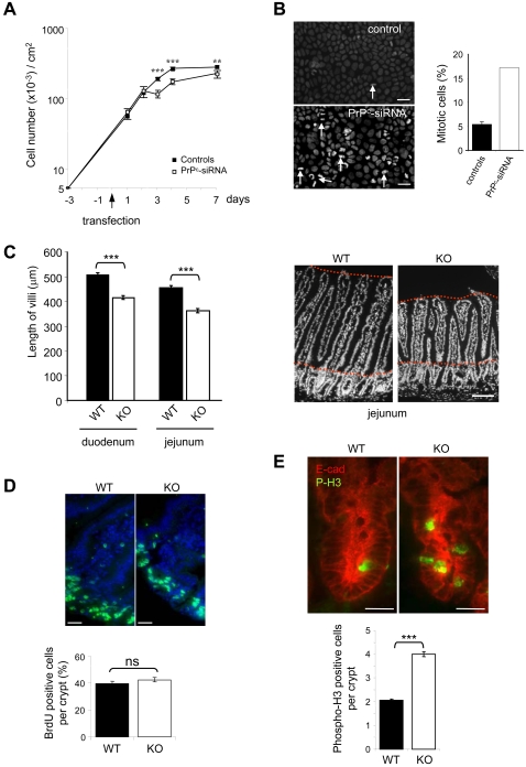
Methodology/findings: We show here that, in cultured human enterocytes and in intestine in vivo, the mature PrP(c) is differentially targeted either to the nucleus in dividing cells or to cell-cell contacts in polarized/differentiated cells. By proteomic analysis, we demonstrate that the junctional PrP(c) interacts with cytoskeleton-associated proteins, such as gamma- and beta-actin, alpha-spectrin, annexin A2, and with the desmosome-associated proteins desmoglein, plakoglobin and desmoplakin. In addition, co-immunoprecipitation experiments revealed complexes associating PrP(c), desmoglein and c-Src in raft domains. Through siRNA strategy, we show that PrP(c) is necessary to complete the process of epithelial cell proliferation and for the sub-cellular distribution of proteins involved in cell architecture and junctions. Moreover, analysis of the architecture of the intestinal epithelium of PrP(c) knock-out mice revealed a net decrease in the size of desmosomal junctions and, without change in the amount of BrdU incorporation, a shortening of the length of intestinal villi.

Conclusions/significance: From these results, PrP(c) could be considered as a new partner involved in the balance between proliferation and polarization/differentiation in epithelial cells.


Conflict of interest statement

Competing Interests: The authors have declared that no competing interests exist.

Figures

Figure 1
Figure 1. Expression and localization of PrPc in proliferating or differentiated/polarized Caco-2/TC7 cells.
Immunofluorescence labeling of PrPc (red, 12F10 antibody) and E-cadherin (green) was performed after 3 (A, left panel) or 10 (D, left panel) days in culture. Nuclei were stained with DAPI (A, middle and right panels, D, right panel). Right panels in A represent an enlargement of a typical nuclear labeling of one cell (*, nucleolus). Note that left panels correspond to a cluster of three cells and middle panels to a cluster of 13 cells. (B): Immunoelectron microscopy of PrPc was performed on day 3. (N, nucleus; *, nucleolus). (C, E): Sub-cellular localisation of GFP-PrPc was analyzed on day 3 or 10 and compared with DAPI staining (right panels). Note the absence of PrPc and of E-cadherin, used as a marker of the junctional state, at the cell–cell contacts of proliferative cells (A) and their presence in the lateral membranes of polarized/differentiated cells as shown in XY (upper panels D) and XZ (lower panel D) planes. Bars: (A) 10 µm for left panels, 20 µm for middle panels and 4 µm for right panels, (B) 1 µm, (C and E) 10 µm and (D) 20 µm.
Figure 2
Figure 2. Expression and localization of PrPc and Ki67 in normal human intestinal epithelium.
Immunofluorescence labeling of PrPc (green, 12F10 antibody) and Ki67 (red) was performed on thin sections of paraffin-embedded pieces of jejunum. Images in A, B and C correspond to crypts, crypt-villus transition compartment and villus respectively, as indicated on the scheme. Lower panels in A correspond to an enlargement of the crypt zone shown above. In B and C, nuclei were stained with DAPI. Note the colocalization of PrPc and Ki67 in the nuclei of crypt cells and the cytoplasmic and membrane localization of PrPc in Ki67 negative epithelial cells. LP, lamina propria; L, lumen. Arrows point out cell–cell junctions of the epithelium. Bars: 20 µm.
Figure 3
Figure 3. Biochemical characterization of membrane- and nucleus-associated PrPc isoforms.
(A): Stability of membrane and nuclear PrPc was analyzed by western blot after treatment of the cells with cycloheximide for the indicated times and purification of membranes and nuclei. Bands obtained in western blots (SAF 32 antibody) were quantified by scanning densitometry. E-cadherin and PARP were used to normalize the values obtained in membrane and nuclear fractions respectively, since both proteins were found stable for the duration of CHX treatment. Histograms correspond to the ratio (%) between PrPc and E-cadherin or PARP from the corresponding scanned bands at each time (mean±SD from 3 independent experiments), the value obtained at time 0 being set at 100. (B): To determine the glycosylation state, rafts and nuclear extracts were treated (+) or not (−) with endo F and PrPc was analyzed by western blot (SAF 32 antibody). Molecular weight in KDa are indicated (C): The presence of a GPI anchor was analyzed after immunoprecipitation of PrPc from rafts or nuclear extracts, SDS-PAGE, transfer and overlay with biotinylated pro-aerolysin bacterial toxin. To check the purity of the extracts, the expression of calnexin (membrane marker) and PARP (nuclear marker) was analyzed by western blot. Molecular weight in KDa are indicated.
Figure 4
Figure 4. Immunodetection of PrPc-associated desmosomal proteins in differentiated Caco-2/TC7 cells.
(A): Proteins that were found to interact with PrPc through proteomic study (Table 1) were analyzed by western blot in rafts, and in immunoprecipitated material (IP) or in supernatants of immunoprecipitations (S-IP) from raft extracts of differentiated cells (day 10). Immunoprecipitations were performed with rabbit anti-PrPc antibodies (Ab703) or with non-specific rabbit immunoglobulins as a control. (B): The presence of E-cadherin, which was not identified as a PrPc partner, was checked, after immunoprecipitation with anti-PrPc, in immunoprecipitated material (IP) and immunoprecipitation supernatant (S-IP). Note that E-cadherin is only recovered in immunoprecipitation supernatant. (C): desmoglein (Dsg), Src, PrPc and E-cadherin (E-cad) were analyzed by western blot in raft extracts (right lane) or after their immunoprecipitation with anti-desmoglein antibodies (left panel). Note the co-immunoprecipitation of desmoglein, c-Src and PrPc and the absence of E-cadherin co-immunoprecipitation.
Figure 5
Figure 5. Effects of PrPc invalidation on the sub-cellular localization of junctional PrPc partners and on desmosome structure.
(A): Caco-2/TC7 cells were transfected by PrPc-siRNA 3 days after seeding and the expression and localization of PrPc (red, 12F10 antibody) and E-cadherin (green) were analyzed 3 days and 7 days after transfection in control (Oligofectamine or mouse PrPc-siRNA transfected cells) or human PrPc-siRNA transfected cells. Phase contrast image (phase) shows the morphology of the cell layer. (B): Immunofluorescence labeling of PrPc (red), c-Src, desmoglein, plakoglobin, desmoplakin and F-actin (green) was performed in control cells (left panels) and in human PrPc-siRNA transfected cells 3 days after transfection (right panels). Control pictures are representative of the results obtained with scramble siRNA or with mouse PrPc si-RNA or with cells incubated with the transfection agent, Oligofectamine. In human PrPc-siRNA transfected cells, fields combining zones where PrPc was still present (open arrowhead) and zones where it was switched off (closed arrowhead) are shown. Note the difference in cell shape between both areas. Mouse 12F10 anti-PrPc antibody was used for immunofluorescence labeling of PrPc except for co-labeling with plakoglobin, in which rabbit polyclonal anti-PrPc Ab703 antibodies were used. Bar: 20 µm. (C): Desmosomes were analyzed by electron microscopy in intestinal epithelium sections from PrPc knock out (KO) and wild type (WT) mice. Note the electron-dense desmosomal plaque which was shorter in PrPc-knock-out than in wild type mice (arrowheads). mv: microvilli, TJ: tight junction, AJ: adherens junction. Bar: 100 nm. Quantification of desmosome size was represented in the lower graph. Box and whisker plots show median desmosomal length (horizontal line), 25th and 75th centiles (box), and range (whiskers) in the wild type (WT, n = 135, three mice) and PrPc-knock out mice (KO, n = 135, three mice), ***p<0.001.
Figure 6
Figure 6. Effects of PrPc invalidation on the completion of cell division and length of intestinal villi.
(A): Caco-2/TC7cells were numbered all along the experiment from 3 days before transfection (−3) to 7 days after transfection. Controls combine results obtained in the three conditions described in figure 5 (scramble siRNA, mouse PrPc si-RNA, Oligofectamine). PrPc-siRNA condition corresponds to the results obtained after specific human PrPc-siRNA transfection. Results are from 4 independent experiments. **p<0.01, ***p<0.001. (B): In the same experiments, cells that showed nuclear division phases were counted after staining with DAPI. In the left panels arrows point examples of metaphases or telophases that were counted. In each condition, 1000 cells were analyzed and the percentage of mitotic cells in controls (black bar, same three conditions as above) or in human PrPc-siRNA (white bar) transfected cells is reported in the right panel. Error bar shown in histogram of controls represents the statistical analyses performed when combining the three control conditions. C: sections of duodenum and jejunum fragments from wild type (WT) and PrPc knock out (KO) mice (2 mice in each group) were stained with DAPI (right panel, bar: 100 µm). Sixty villi were measured for each mouse and fragment. No difference was observed between the 2 mice of each group (not shown), but significant differences (***p<0.001) were obtained between wild type (black bars) and knock out mice (white bars) in both duodenum and jejunum fragments. (D): After 1.5 hour BrdU intraperitoneal injection, sections from the jejunum fragment of wild type (WT) and knock out (KO) mice were performed. Nuclei were visualized with DAPI staining (blue). BrdU-stained proliferating cells (green) are limited to the crypts in wild type and knock out animals. Bars: 10 µm. Knock out mice display similar number of positive BrdU cells per crypt as compared with wild type mice (quantification in the right panel graph). No significant difference was revealed by statistical analysis (ns, student's t test). (E): Immunofluorescence analyses of phospho H3 (green) and E-cadherin (red) were performed on intestine sections from wild type (WT) or PrPc knock out (KO) mice (3 mice in each group). Pictures of crypt staining are shown (upper panels; bars: 10 µm) and the number of phospho-H3 positive cells per crypt is reported (155 crypts were counted in each group). ***p<0.001.

Similar articles

Cited by

References

    1. Prusiner SB, Scott MR, DeArmond SJ, Cohen FE. Prion protein biology. Cell. 1998;93:337–348. - PubMed
    1. Bueler H, Aguzzi A, Sailer A, Greiner RA, Autenried P, et al. Mice devoid of PrP are resistant to scrapie. Cell. 1993;73:1339–1347. - PubMed
    1. Prusiner SB. The prion diseases. Brain Pathol. 1998;8:499–513. - PMC - PubMed
    1. Weissmann C, Bueler H, Fischer M, Sailer A, Aguzzi A, et al. PrP-deficient mice are resistant to scrapie. Ann N Y Acad Sci. 1994;724:235–240. - PubMed
    1. Bendheim PE, Brown HR, Rudelli RD, Scala LJ, Goller NL, et al. Nearly ubiquitous tissue distribution of the scrapie agent precursor protein. Neurology. 1992;42:149–156. - PubMed

Publication types