Comparative Study
. 2008 Sep;10(9):964-72.
doi: 10.1593/neo.08604.

Clusterin interacts with Paclitaxel and confer Paclitaxel resistance in ovarian cancer

Affiliations
Comparative Study

Clusterin interacts with Paclitaxel and confer Paclitaxel resistance in ovarian cancer

Dong Choon Park et al. Neoplasia. 2008 Sep.

Abstract

Optimal debulking followed by chemotherapy is the standard treatment of managing late-stage ovarian cancer, but chemoresistance is still a major problem. In this study, we compared expression profiles of primary tumor tissue from five long-term (>8 years) and five short-term (<2 years) ovarian cancer survivors and identified clusterin as one of the genes that were significantly up-regulated in short-term survivors. We then evaluated the prognostic significance of clusterin and its possible correlation with chemoresistance in ovarian cancer by immunohistostaining of clusterin in 62 tumor samples from patients with stage III, high-grade serous ovarian cancer. After adjusting for debulking status and age, Cox regression analyses showed that high levels of clusterin expression correlate with poor survival (hazard ratio, 1.07; 95% confidence interval, 1.002-1.443; P = .04). We also investigated clusterin in paclitaxel resistance by modulating the endogenous clusterin expression in ovarian cancer cells and treating the cells with purified clusterin. Results indicate that high-clusterin-expressing ovarian cancer cells are more resistant to paclitaxel. Moreover, exposing ovarian cancer cells to exogenous clusterin increases cells' resistance to paclitaxel. Finally, using size exclusion chromatography and fluorescently labeled paclitaxel, we demonstrated that clusterin binds to paclitaxel. In summary, our findings suggest that high levels of clusterin expression increase paclitaxel resistance in ovarian cancer cells by physically binding to paclitaxel, which may prevent paclitaxel from interacting with microtubules to induce apoptosis. Thus, clusterin is a potential therapeutic target for enhancing chemoresponsiveness in patients with a high-level clusterin expression.


Figures
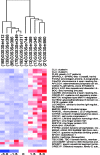
Figure 1
Figure 1
Partial list of differentially expressed genes in tumors from long- and short-term ovarian cancer survivors. The length of survival for each patient is shown within a labeled bracket. Red, white, and blue indicate a fold-change expression level above, at, and below the mean expression of a gene across all samples, respectively.
Figure 2
Figure 2
Clusterin expression correlates with overall survival and chemoresponse. (A) Kaplan-Meier survival curves for patients with stage III, high-grade serous ovarian adenocarcinomas. Patients who had tumors with positive clusterin expression (+) had significantly poorer prognoses than those who had negative clusterin expression (-), whether their debulking was optimal (i.e., the largest residual tumor was <2 cm) or suboptimal (i.e., the largest residual tumor was >2 cm). (B) Box-plot showing clusterin protein expression in chemoresponders and nonresponders. The box is bounded above and below by the 75th and 25th percentiles, and the median is the line in the box. Whiskers are drawn to the nearest value not beyond a standard span from the quartiles; points beyond (outliers) are drawn individually, where the standard span is 1.5 x (interquartile range).
Figure 3
Figure 3
Correlation of clusterin expression and paclitaxel resistance. (A) Western blot analysis of clusterin expression in 13 epithelial ovarian cancer cell lines. (B) Scatter plot showing correlation between clusterin expression and paclitaxel IC50 for the 13 ovarian cancer cell lines tested. High IC50 values denote paclitaxel resistance, whereas low IC50 values indicate paclitaxel sensitivity. A Spearman correlation test showed that clusterin overexpression significantly correlated with IC50 for paclitaxel (R2 = 0.55, P = .004).
Figure 4
Figure 4
Increased paclitaxel resistance in clusterin transfectants. (A) Western blot analysis showing overexpression of clusterin in SKOV3 cells stably transfected with plasmid pcDNA3.1 with clusterin (cDNA3.1-Clu). (B) SKOV3-cDNA3.1-Clu stably transfected expressing clusterin had higher survival rates than the control cells SKOV3-vector, which were transfected with pcDNA3.1 vector only.
Figure 5
Figure 5
Exogenous clusterin enhances the paclitaxel resistance of SKOV3 cells. Cell proliferation was measured by XTT assay in SKOV3 cells treated with 10-7 M paclitaxel (T) for 48 hours in the presence of the indicated concentrations of clusterin. The baseline used for calculating relative cell proliferation rate was set by adjusting the proliferation rate of paclitaxel-treated SKOV3 cells without exogenous clusterin to 1.
Figure 6
Figure 6
PEO4 became sensitive to paclitaxel after silencing clusterin expression with siRNA. (A) Western blot analyses show the time course of clusterin silencing in PEO4 cells transfected with siRNA for clusterin (+) or luciferase (-). After transfection, the cells were cultured for 2 days in medium containing 10-7 M paclitaxel. (B) Cell survival after paclitaxel treatment as determined by XTT assay.
Figure 7
Figure 7
Effects of decreased clusterin expression on paclitaxel resistance in xenografted tumors. (A) When xenografts in nude mice resulting from the inoculation of paclitaxel-resistant cells transduced with shRNA for clusterin or the Lacz gene grew to approximately 100 mm3, mice were injected with 0.2 mg/0.1 ml per 10 g of paclitaxel intraperitoneally. Tumor volumes were measured twice every week. (B) Western blot analysis of clusterin expression from xenografted tumors from mouse autopsies. Tumors from three separate mice from both the shRNA-Lacz and shRNA-clusterin groups were analyzed.
Figure 8
Figure 8
Exogenous clusterin prevents the binding of paclitaxel to microtubules in SKOV3 cells. PEO4 (A and B) and SKOV3 (C and D) cells cultured in chamber coverglasses were treated with 10-6 M OG-labeled paclitaxel in the absence (A and C) or presence (B and D) of 7.5 µg/ml clusterin for 30 minutes. Cells were also simultaneously incubated with the Hoechst stain for nuclei visualization, and fluorescent images were captured by an inverted fluorescence microscope equipped with a digital camera using the same setting. The binding of OG-labeled paclitaxel to microtubules is shown in panel C.
Figure 9
Figure 9
Coelution of clusterin and OG-labeled paclitaxel (paclitaxel- OG) in size exclusion chromatography. Solutions containing clusterin and paclitaxel-OG mixture, paclitaxel-OG alone, or a GST and paclitaxel-OG mixture (all at 5 mM) were fractionated by size exclusion chromatography in PEM buffer, and the OG fluorescence and A280 of the eluate were measured. The upper panel shows OG fluorescence as a function of elution time; the lower panel shows the corresponding A280 profile. The result shown is representative of three independent experiments.

References

    1. Boring CC, Squires TS, Tong T, Montgomery S. Cancer statistics, 1994. CA Cancer J Clin. 1994;44:7–26. - PubMed
    1. Landis SH, Murray T, Bolden S, Wingo PA. Cancer statistics, 1998. CA Cancer J Clin. 1998;48:6–29. - PubMed
    1. Omura GA, Brady MF, Homesley HD, Yordan E, Major FJ, Buchsbaum HJ, Park RC. Long-term follow-up and prognostic factor analysis in advanced ovarian carcinoma: the Gynecologic Oncology Group experience. J Clin Oncol. 1991;9:1138–1150. - PubMed
    1. Barlund M, Forozan F, Kononen J, Bubendorf L, Chen Y, Bittner ML, Torhorst J, Haas P, Bucher C, Sauter G, et al. Detecting activation of ribosomal protein S6 kinase by complementary DNA and tissue microarray analysis. J Natl Cancer Inst. 2000;92:1252–1259. - PubMed
    1. Wang T, Hopkins D, Schmidt C, Silva S, Houghton R, Takita H, Repasky E, Reed SG. Identification of genes differentially over-expressed in lung squamous cell carcinoma using combination of cDNA subtraction and microarray analysis. Oncogene. 2000;19:1519–1528. - PubMed

Publication types

LinkOut - more resources