. 2008 Oct;21(5):565-78.
doi: 10.1111/j.1755-148X.2008.00482.x. Epub 2007 Jun 28.

Iris phenotypes and pigment dispersion caused by genes influencing pigmentation

Affiliations

Iris phenotypes and pigment dispersion caused by genes influencing pigmentation

Michael G Anderson et al. Pigment Cell Melanoma Res. 2008 Oct.

Abstract

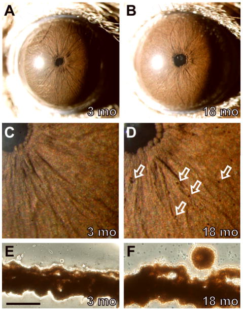
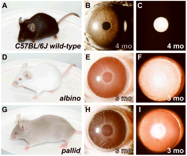
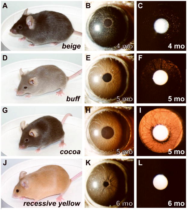
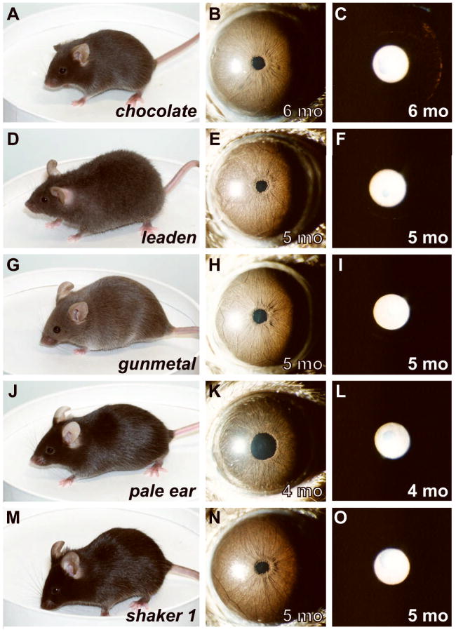
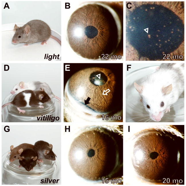
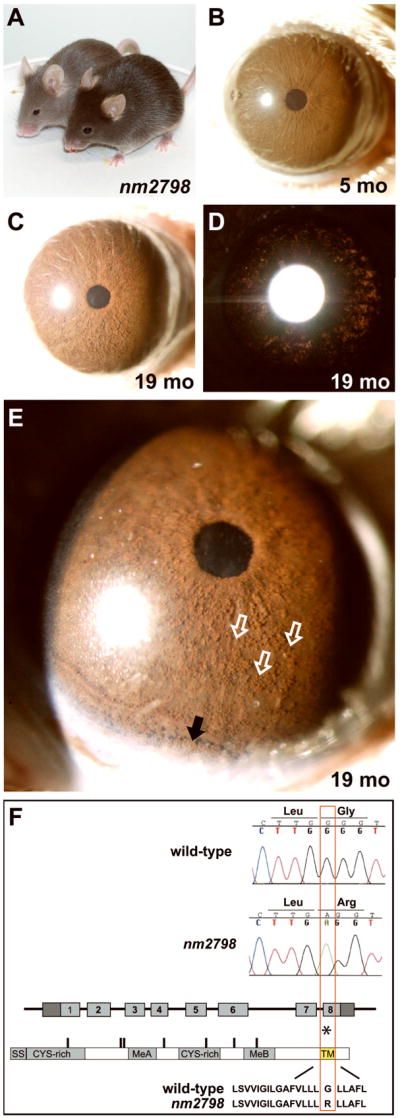
Spontaneous mutations altering mouse coat colors have been a classic resource for discovery of numerous molecular pathways. Although often overlooked, the mouse iris is also densely pigmented and easily observed, thus representing a similarly powerful opportunity for studying pigment cell biology. Here, we present an analysis of iris phenotypes among 16 mouse strains with mutations influencing melanosomes. Many of these strains exhibit biologically and medically relevant phenotypes, including pigment dispersion, a common feature of several human ocular diseases. Pigment dispersion was identified in several strains with mutant alleles known to influence melanosomes, including beige, light, and vitiligo. Pigment dispersion was also detected in the recently arising spontaneous coat color variant, nm2798. We have identified the nm2798 mutation as a missense mutation in the Dct gene, an identical re-occurrence of the slaty light mutation. These results suggest that dysregulated events of melanosomes can be potent contributors to the pigment dispersion phenotype. Combined, these findings illustrate the utility of studying iris phenotypes as a means of discovering new pathways, and re-linking old ones, to processes of pigmented cells in health and disease.


Figures

Figure 1
Figure 1
Normal iris phenotypes of wild-type C57BL/6J mice. Comparisons of a representative 3-mo eye (left column) to an 18-mo eye (right column). (A) The normal C57BL/6J iris of young mice as viewed with broadbeam illumination originally imaged at 25X magnification. The iris is characterized by a sienna-brown color, a complex surface morphology with several small underlying vessels, and a circular pupil. The bright white circle to the left of the pupil is a reflection from flash photography. (B) With age, a number of clump cells are present on the surface of the iris and the iris becomes slightly more reddish in color. (C,D) At higher 40X magnification and less image reduction, clump cells are more readily visible, each casting a characteristic small crescent shadow (several are indicated by open white arrows, others in the field are unmarked). (E,F) Unstained cryosections of the same eyes shown above showing the presence of a melanin engulfed phagocytic clump cell in panel F on the surface of the iris stroma as a normal consequence of aging. These cells were not visible in 0/64 sections from the young eye in E and were present on the iris in 6/70 sections analyzed from the aged eye in F. Scale bar = 50 μm.
Figure 2
Figure 2
Substrains exhibiting a correlation between coat and iris appearance. Coat color (left column), broadbeam illumination of iris (middle column), and transilluminating view of iris (right column). The bright white circle (to the left of the pupil with broadbeam illumination and central with transilluminating illumination) is a reflection from flash photography. (A–C) Wild-type C57BL/6J mice have darkly pigmented coats and darkly pigmented irides that are sienna-brown in color. Because the irides are darkly pigmented, the iris appears black with transilluminating illumination. (D–E) albino mice have coats and irides totally lacking melanin pigment. (G–I) pallid mice have severely diluted coats and irides. All mice were homozygous for the indicated mutations and were maintained on a C57BL/6J genetic background.
Figure 3
Figure 3
Substrains exhibiting discordant coat color and iris appearances. Coat color (left column), broadbeam illumination of iris (middle column), and transilluminating view of iris (right column). (A–C) beige mice have slightly diluted coats, but irides that appear very dark. (D–F) buff mice have relatively severe dilution of coat color, but only a slight dilution of iris pigmentation evident by mild transillumination defects. The transillumination defects exhibited no discernable pattern, other than that the extent of the defects tended to increase peripherally. No influence of age was detectable. (G–I) cocoa mice have a modest dilution of coat color but strikingly hypopigmented irides (J–L) recessive yellow mice have yellow coats, but normally pigmented irides that are indistinguishable from those of normally pigmented C57BL/6J mice. All mice were homozygous for the indicated mutations and were maintained on a C57BL/6J genetic background.
Figure 4
Figure 4
Substrains with mild, or no, change in iris appearance. Coat color (left column), broadbeam illumination of iris (middle column), and transilluminating view of iris (right column). (A–C) chocolate mice have a modest alteration in coat color and mild transillumination defects that tended to increase peripherally. These transillumination defects did not worsen with increasing age among the mice examined. In contrast, other strains with similarly modest alterations in coat color maintain completely normal irides; including, (D–F) leaden mice have slightly diluted coats and normal appearing irides (the leaden substrain utilized here also carried the fuzzy allele, so it can additionally be concluded that fuzzy does not influence the iris), and (G–I) gunmetal mice have slightly diluted coats and normal appearing irides. Two substrains with mutations that do not influence adult coat color at all, but do influence melanosomes in other tissues were also examined; including, (J–L) As adults, pale ear mice have normal coat color but irides that appear slightly darker (note that the ears and tail of pale ear mice are also light colored), and (M–O) shaker 1 mice have normally colored coats and normal appearing irides. All mice were homozygous for the indicated mutations and were maintained on a C57BL/6J genetic background.
Figure 5
Figure 5
Pigment dispersion is a common, but not universal, feature of mouse strains with mutations related to Tyrp1. Comparisons of coat color (left column) and ocular phenotypes (right columns). (A) The light allele in LT/SvEiJ mice results in the production of hair that is pigmented at the tip, but very lightly or not pigmented along the hair shaft. As a consequence, coat color is rapidly lightened as hair lengthens. (B) Pigment dispersion was commonly present in the eyes of aged light mice. (C) Higher magnification image of same pupil shown in panel B. Several clumps of dispersed pigment are clearly visible in the pupil (one prominent example indicated with arrowhead, several other unmarked also visible). (D) The vitiligo allele results in a coat that is initially lighter than normal, with extensive white spotting (front mouse). With increasing age, the coat becomes progressively whiter due to increasing numbers of white hairs with each molt (back mouse). (E) Pigment dispersion was striking in eyes of aged vitiligo mice, as shown here in the pupil (arrowhead), across the surface of the iris (open white arrow), and in a pronounced pool accumulated inferiorly (dark band marked by solid black arrow). (F) Eyes of several vitiligo mice became severely enlarged, as sometimes occurs in mice with glaucoma. (G) The silver allele results in a mix of normal and hypopigmented hairs, which together cause a characteristic silvering of the coat. Heterozygosity for Tyrp1b enhances the silver coat color phenotype as shown here among three young silver homozygotes with differing Tyrp1 genotypes (Tyrp1+/+ front, Tyrp1b/b left, and Tyrp1b/+ right). (H) Mice heterozygous for Tyrp1b and homozygous for Sisi maintain healthy irides lacking pigment dispersion. (I) Mice homozygous for both Tyrp1b and Sisi exhibit a characteristic Tyrp1 mutant iris phenotype characterized by iris atrophy and pigment dispersion. Neither the onset nor severity of these Tyrp1b mediated phenotypes were influenced by Si. Note, because of the unique photographic flash settings used to capture the pigment accumulation near the highly reflective sclera in panel E, the apparent color of this iris is not directly comparable to the images in other panels.
Figure 6
Figure 6. Phenotypes and sequence analysis of the spontaneously occurring nm2798 mutation
(A) The nm2798 mutation acts semi-dominantly to influence coat color, as shown here in a comparison of a heterozygote (front) and homozygote (back). (B) Healthy iris of a young nm2798 homozygote. (C) Aged nm2798 homozygote showing dispersed pigment across the surface of the iris and (D) mild transillumination defects spread across the iris. (E) Higher magnification view with a narrower slit of illumination showing the same iris as in panels C,D. The discretely rounded appearance of the dispersed pigment along the iris surface (three examples marked with open white arrows, several others in field unmarked) indicates that it is likely present within phagocytic clump cells. Pigment is also accumulating inferiorly (solid black arrow). (F) Sequence comparison of wild-type (B6) versus mutant (nm2798) alleles. A single base pair substitution within exon 8 of the Dct gene results in a mis-sense mutation changing a glycine to arginine (asterisk). The base pair and amino acid changes are both identical to previously described slaty light mutation. The change occurs within the predicted transmembrane spanning domain (TM). Other motifs of the DCT protein include a signal sequence (SS); cysteine rich domains (CYS-rich); and metal-binding motifs (MeA, MeB).

References

    1. Aksan I, Goding CR. Targeting the microphthalmia basic helix-loop-helix-leucine zipper transcription factor to a subset of E-box elements in vitro and in vivo. Mol Cell Biol. 1998;18:6930–8. - PMC - PubMed
    1. Anderson MG, Libby RT, Mao M, Cosma IM, Wilson LA, Smith RS, John SW. Genetic context determines susceptibility to intraocular pressure elevation in a mouse pigmentary glaucoma. BMC Biol. 2006;4:20. - PMC - PubMed
    1. Anderson MG, Smith RS, Hawes NL, Zabaleta A, Chang B, Wiggs JL, John SW. Mutations in genes encoding melanosomal proteins cause pigmentary glaucoma in DBA/2J mice. Nat Genet. 2002;30:81–5. - PubMed
    1. Anderson MG, Smith RS, Savinova OV, Hawes NL, Chang B, Zabaleta A, Wilpan R, Heckenlively JR, Davisson M, John SW. Genetic modification of glaucoma associated phenotypes between AKXD-28/Ty and DBA/2J mice. BMC Genet. 2001;2:1. - PMC - PubMed
    1. Ball SF. Pigmentary Glaucoma. In: Yanoff M, Duker JS, editors. Ophthalmology. 2. St Louis: Mosby; 2004.

Publication types