Comparative Study
. 2008 Aug 20:9:392.
doi: 10.1186/1471-2164-9-392.

Rapid functional diversification in the structurally conserved ELAV family of neuronal RNA binding proteins

Affiliations
Comparative Study

Rapid functional diversification in the structurally conserved ELAV family of neuronal RNA binding proteins

Marie-Laure Samson. BMC Genomics. .

Abstract

Background: The Drosophila gene embryonic lethal abnormal visual system (elav) is the prototype of a gene family present in all metazoans. Its members encode structurally conserved neuronal proteins with three RNA Recognition Motifs (RRM) but they paradoxically act at diverse levels of post-transcriptional regulation. In an attempt to understand the history of this family, we searched for orthologs in eleven completely sequenced genomes, including those of humans, D. melanogaster and C. elegans, for which cDNAs are available.

Results: We analyzed 23 orthologs/paralogs of elav, and found evidence of gain/loss of gene copy number. For one set of genes, including elav itself, the coding sequences are free of introns and their products most resemble ELAV. The remaining genes show remarkable conservation of their exon organization, and their products most resemble FNE and RBP9, proteins encoded by the two elav paralogs of Drosophila. Remarkably, three of the conserved exon junctions are both close to structural elements, involved respectively in protein-RNA interactions and in the regulation of sub-cellular localization, and in the vicinity of diverse sequence variations.

Conclusion: The data indicate that the essential elav gene of Drosophila is newly emerged, restricted to dipterans and of retrotransposed origin. We propose that the conserved exon junctions constitute potential sites for sequence/function modifications, and that RRM binding proteins, whose function relies upon plastic RNA-protein interactions, may have played an important role in brain evolution.


Figures

Figure 1
Figure 1
Correspondance between exons and protein regions in the elav family of D. melanogaster. A: RNA structures. RNA nomenclature as in FlyBase, with details in the Methods. Boxes represent exons. The black horizontal lines are introns, with dashes respectively replacing the 5.8 kb long intron in the rbp9-RA transcript and the 2.2 kb long intron in the elav-RA transcript. White: non coding, Vertical stripes: non-conserved, Crossed: gene-specific mini-exons, respectively a 15 nucleotide long region present in alternative forms of rbp9 and a 45 nucleotide long region present in fne. All others are color coded based upon sequence similarity and according to exon-exon boundaries. B: Schematic representation of the ELAV family protein products. The color coding corresponds to that used for the RNA representation. The regions encoded by gene specific sequences have been omitted. RRM: RNA Recogntion Motif. The pairs of white vertical bars represent conserved motifs (RNP-1 and RNP-2) diagnostic of RRMs.
Figure 2
Figure 2
Exon organisation of the elav-related genes in 11 metazoans. The analyzed species are listed on the left, with classical phylogenetic relationships represented. The number of elav-like genes is listed next to the species names. Percentages of identity between their protein products and the D. melanogaster proteins ELAV, FNE and RBP9 are listed on the right side of the figure. At the top, a typical ELAV-like protein is represented, with its three RRMs and the hinge region between RRM2 and 3. The vertical arrows below point at protein regions that are, depending upon each of the 23 analyzed proteins, either encoded by exon-junctions (Jx, x = 1 to 8, see text) or by an internal exon sequence. The presence of the junction-encoded region is indicated by a vertical bar for each protein.
Figure 3
Figure 3
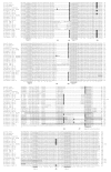
Protein sequence comparison among 27 ELAV-like proteins forms. Alternative protein forms are included, specifically for Drosophila RBP9 (A and D) and three of the human proteins (HuB, HuC and HuD, where HuX-n refers to the n amino acid long form of the HuX protein). "*" indicate that amino acids are identical in all 27 sequences, ":" and "." respectively indicate conserved and semi-conserved substitutions. The octamer RNP-1 and the hexamer RNP-2, diagnostic of RRMs, are underlined. Also underlined is a conserved octamer present in the region that is crucial for nuclear export and localization. The regions in light grey boxes have been mapped as necessary for these processes in D. melanogaster ELAV, human HuR and human HuD. We identified eight exon junctions labelled J1 to J8 (see text). Bold characters and dark grey boxes are used to identify amino acids encoded by exon junctions. When the splicing connects intact codons, two amino acids are bold (J3 and J8). The symbol//replaces 85 non-conserved amino acids in the C. elegans sequence.
Figure 4
Figure 4
Phylogenetic tree of 27 ELAV-like proteins. Sequences were aligned and bootstrapped 500 times. Numbers near the branches are the bootstrap values, and the scale indicates the number of substitutions per site.
Figure 5
Figure 5
A unique nested gene arrangement for the elav and arginase genes in D. melanogaster. A: The elav gene is nested inside the third intron of the arginase gene. Complementary strands are transcribed to generate the elav and arg RNAs with inverse polarities [28]. B: Examination of the relative arg-elav arrangement in 11 metazoans. There are two arginase genes in humans, only one in the other examined species. Column 1 documents the status of the arginase third intron. Column 2 specifies the nested (+) or independent (-) arrangement of the arginase/elav genes. N.A.: Not applicable. The third column indicates the percentage of amino-acid sequence identity of D. melanogaster compared with other species. *: N-terminally truncated arginase sequence for P. humanus corporis. See Additional file 3 for arginase alignments.

References

    1. Yao KM, Samson ML, Reeves R, White K. Gene elav of Drosophila melanogaster: a prototype for neuronal-specific RNA binding protein gene family that is conserved in flies and humans. J Neurobiol. 1993;24:723–739. doi: 10.1002/neu.480240604. - DOI - PubMed
    1. Pascale A, Amadio M, Quattrone A. Defining a neuron: neuronal ELAV proteins. Cell Mol Life Sci. 2008;65:128–140. doi: 10.1007/s00018-007-7017-y. - DOI - PMC - PubMed
    1. Keene JD. Why is Hu where? Shuttling of early-response-gene messenger RNA subsets. Proc Natl Acad USA. 1999;96:5–7. doi: 10.1073/pnas.96.1.5. - DOI - PMC - PubMed
    1. Lunde BM, Moore C, Varani G. RNA-binding proteins: modular design for efficient function. Nat Rev Mol Cell Biol. 2007;8:479–490. doi: 10.1038/nrm2178. - DOI - PMC - PubMed
    1. Maris C, Dominguez C, Allain FH. The RNA recognition motif, a plastic RNA-binding platform to regulate post-transcriptional gene expression. FEBS J. 2005;272:2118–2131. doi: 10.1111/j.1742-4658.2005.04653.x. - DOI - PubMed

Publication types

LinkOut - more resources