. 2008 Nov;82(21):10820-31.
doi: 10.1128/JVI.00365-08. Epub 2008 Aug 20.

Natural killer cells as novel helpers in anti-herpes simplex virus immune response

Affiliations

Natural killer cells as novel helpers in anti-herpes simplex virus immune response

Subhadra Nandakumar et al. J Virol. 2008 Nov.

Abstract

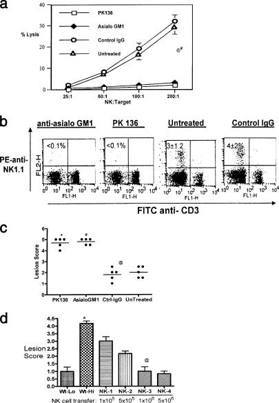
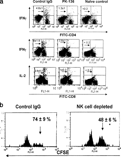
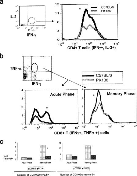
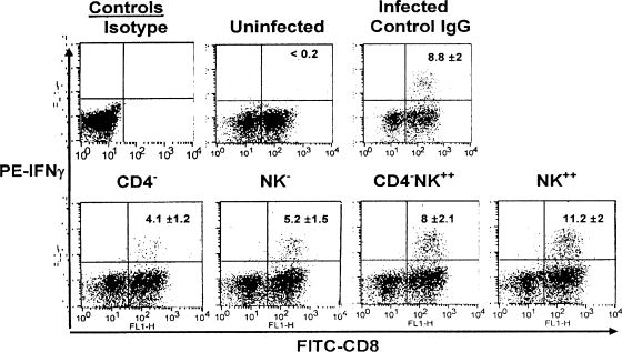
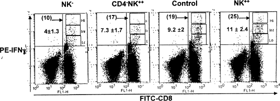
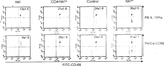
Innate defenses help to eliminate infection, but some of them also play a major role in shaping the magnitude and efficacy of the adaptive immune response. With regard to influencing subsequent adaptive immunity, NK cells aided by dendritic cells may be the most relevant components of the innate reaction to herpes simplex virus (HSV) infection. We confirm that mice lacking or depleted of NK cells are susceptible to HSV-induced lesions. The quantity and quality of CD8(+) cytotoxic T lymphocytes generated in the absence of NK cells were diminished, thereby contributing to susceptibility to HSV-induced encephalitis. We demonstrate a novel helper role for NK cells, in that NK cells compensate for the loss of CD4 helper T cells and NK cell supplementation enhances the function of wild type anti-HSV CD8 T cells. In addition, NK cells were able to partially rescue the dysfunctional CD8(+) T cells generated in the absence of CD4 T helper cells, thereby performing a novel rescue function. Hence, NK cells may well be exploited for enhancing and rescuing the T-cell response in situations where the CD4 helper response is affected.


Figures

FIG. 1.
FIG. 1.
Lack of NK cells contributes to susceptibility, while supplementation enhances resistance. (a) Mice given anti-asialo-GM1 and anti-NK 1.1 antibodies lack detectable NK cell lytic activity. Sample animals from groups administered anti-asialo-GM1 or control IgG were sacrificed 48 h later. Spleen cells were analyzed for in vitro NK cell cytotoxic activity. A suspension of 107/ml of spleen cells was serially diluted with 100 ml in each triplicate well of a 96-well round-bottomed plate. Target 51Cr-labeled YAC-1 mouse lymphoma cells were added to give a effector/target cell ratio of 200, 100, 50, or 25. After 4 h of incubation at 37°C, supernatants containing released 51Cr were collected and counted with an automatic scintillation counter. Specific lysis was calculated as (experimental release − spontaneous release)/(total release − spontaneous release) × 100%, where spontaneous release was derived from wells without effectors and total release from wells with 3% Triton-X added to it. @, asialo-GM1 versus control IgG or untreated, P < 0.0001; #, PK136 versus control IgG and untreated controls, P < 0.0001. (b) Depletion of NK cells by anti-NK1.1 and asialo-GM1 antibodies. B6 mice given anti-asialo-GM1 and PK136 were analyzed for the absence of NK cells by flow cytometry. Single-cell suspensions of spleens were stained with anti-NK1.1 (PE) and anti-CD3 (FITC). The plots represent data obtained from one mouse in each group. The numbers within the plot are the means ± SDs for five mice in each group. The controls included untreated mice and mice given control IgG. Cells that are CD3 NK1.1+ are taken to be the NK cells. (c) Early onset of lesions upon zosteriform challenge in NK-depleted mice. Zoster challenge experiments were performed as described elsewhere (20). Before challenge, the left flank area was depilated by a combination of hair clipping and use of the chemical Nair (Carter-Wallace, New York, NY). The animals were anesthetized with avertin, and scarifications were made in a ∼4-mm2 area. To such scarifications, 10 μl containing 104 PFU of HSV-1 (strain 17) was added and gently massaged. Animals were inspected daily for the development of zosteriform ipsilateral lesions, general behavior changes, encephalitis, and mortality. The severity of the lesions was scored as follows: 1+, vesicle formation; 2+, local erosion and ulceration of the local lesion; 3+, mild to moderate ulceration; 4+, severe ulceration, hind limb paralysis, and encephalitis; and 5+, ultimate death (*, mice that were moribund and hence euthanized). The experiments were repeated three times with five mice in each group, and the outcomes were similar. The lesion scores of all mice within a group at day 10 postchallenge from one such experiment are shown. @, Asialo-GM1 versus control IgG and untreated, P < 0.0001; #, PK136 versus control IgG and untreated, P < 0.0001. (d) NK cell transfer augments protection against HSV. Wild-type B6 mice were divided into six groups of three mice each. Groups 1 and 2 were challenged (zosteriform) with low (5 × 10³) (Wt-Lo) and high (1 × 10⁵) (Wt-Hi) doses of virus, respectively. Groups 3 to 6 (NK-1, -2, -3, and -4, respectively) were adoptively transferred with increasing numbers of purified NK cells as indicated 15 h before challenge with the high dose of virus. The animals were handled as described for panel c and lesion scores recorded. The experiment was repeated three times with three mice per group. Data collected on day 10 postchallenge in one such experiment are shown. *, animals in this group were moribund and hence sacrificed. @, statistically significant difference between NK-3 (1 million NK cells) and Wt-Hi (P < 0.05).
FIG. 2.
FIG. 2.
NK cells contribute to skewing toward the protective Th1 type. (a) Reduced Th1 cytokine production in anti-HSV CD4 and CD8 T cells. C57BL/6 mice were divided into two groups. One group was depleted of NK cells; the other group was administered control IgG and was infected with HSV by the footpad route. At 10 days p.i., their splenocytes were harvested and stimulated with UV-inactivated HSV or SSIEFARL peptide and analyzed for the CD4 and CD8 T-cell activity. Samples were processed individually, and similar patterns were observed within the same group. IFN-γ-producing CD4+ T cells and IFN-γ- and IL-2-producing CD8+ T cells in the control IgG, NK-depleted, and uninfected control animals are shown. The experiment was repeated three times with similar outcomes. The dot plot shows results obtained from one such experiment, and the numbers within the plot are the means ± SDs for five mice. (b) In vivo cytolytic ability is reduced in NK-depleted mice. Splenocytes from wild-type B6 mice were stained with CFSE and loaded with SSIEFARL peptide. A total of 5 × 10⁶ cells were adoptively transferred through the tail vein into control mice and NK-depleted mice. The spleens were isolated from these mice after 5 h and the splenocytes isolated. Target cells were distinguished from recipient cells based on CFSE staining. Histogram plots were used to demonstrate the difference in separation pattern based on intensity of CFSE staining. The recovery and percent killing of the various CFSE-labeled, peptide-pulsed targets were calculated as follows: 100 − ([(percentage of peptide pulsed in immunized mice/percentage of peptide unpulsed in immunized mice)/(percentage of peptide pulsed in unimmunized mice/percentage of peptide unpulsed in unimmunized mice)] × 100). The experiment was repeated two times with three mice in each group. The histogram plot shows the data obtained from one such experiment, and the number within the plot is the mean ± SD of killing ability for three individual mice.
FIG. 3.
FIG. 3.
Reduced frequency and diminished quality of anti-HSV specific CD4+ T helper and CD8+ T-cell numbers in PK136 Tg mice. C57BL/6 mice and PK136 mice were infected with HSV-1 strain 17 by the skin route and analyzed for CD4+ and CD8+ T-cell activity at 7 days p.i. to study the primary immune response and at 45 days p.i. to study the memory response. Splenocytes were harvested at the indicated time points and analyzed for polyfunctional CD4+ T cells (IFN-γ+ and IL-2+) and CD8+ T cells (IFN-γ+ and TNF-α+). In addition, the cytolytic ability of CD8+ T cells was assessed by measuring granzymes and degranulation (CD107a/b). The experiment was done multiple times with similar outcomes. The data shown are representation of one such experiment. (a) Reduced number of polyfunctional CD4+ T cells during the memory phase of infection. Single-cell suspensions were processed as described in Materials and Methods. The sample was gated on IL-2 and IFN-γ double-positive cells and analyzed for CD4 expression. The histogram plot shows the number of CD4+ T cells that are IFN-γ+ and IL-2+ in C57BL/6 and PK136 Tg mice. The patterns were similar in all of the mice within the group. *, the difference is statistically significant. (b) Reduced number of anti-HSV polyfunctional CD8+ T cells during acute and memory infection in PK136 Tg mice. Single-cell suspensions was stained as described above. The cells were gated on IFN-γ+ and TNF-α+ cells as indicated and analyzed for HSV gB tetramer positivity. A representative plot showing the number of gB tetramer-positive CD8+ T cells producing both IFN-γ and TNF-α (polyfunctional CD8+ T cells) during the primary and memory immune responses is shown. *, the difference between the wild-type and PK136 Tg mice was significant. (c) PK136 Tg mice develop reduced CD8+ T cells with cytotoxic ability in HSV infection. Single-cell suspensions were processed as described in Materials and Methods. The analysis for cytolytic activity was done on the CD8+ HSV gB tetramer-positive gated population. A pair of graphs representing the CD107 a/b (left)- and granzyme (right)-expressing HSV-gB tetramer-positive CD8+ T cells in PK136 Tg mice and wild-type mice during primary and recall immune responses is shown. *, although the response was lower in PK136 Tg mice, the difference was statistically significant only during the memory phase analysis.
FIG. 4.
FIG. 4.
NK cells affect CD8+ T-cell IFN-γ production, but addition can compensate for the loss of CD4+ T-cell help. C57BL/6 mice with HSV memory were divided into five groups: (i) depleted of NK cells (NK), (ii) depleted of CD4+ T cells (CD4), (iii) depleted of CD4+ T cells but supplemented with NK (CD4 NK++), (iv) supplemented with NK cells (NK++), and (v) receiving control IgG. All the mice were given 1 × 10⁵ PFU of HSV and 5 days later were analyzed for IFN-γ production by CD8+ T cells to measure recall responses. The patterns were similar in all three experiments. A representative plot of intracellular IFN-γ staining in peptide-stimulated CD8+ T cells is shown. The data represents one mouse from each of the following groups: uninfected control, control IgG, CD4, NK, CD4 NK++, and NK++. The number within the plot is the mean ± SD for five mice in each group. The differences between the NK++ group and the CD4 and NK groups were statistically significant (P < 0.005). The difference between the NK++ and CD4 NK++ groups was significant (P < 0.01).
FIG. 5.
FIG. 5.
NK cells contribute to higher IFN-γ production by anti-HSV CD8+ T cells. As described for Fig. 4, C57BL/6 HSV memory mice were divided into four groups, in which mice from one group were depleted of NK cells (NK), another was depleted of CD4 T cells and supplemented with NK cells (CD4 NK++), the third was supplemented with NK cells (NK++), and the fourth served as a control and received control IgG. All the mice were given 1 × 10⁵ PFU of HSV and 5 days later analyzed for IFN-γ production. Representative dot plots show the CD8 T cells producing IFN-γ in the NK-depleted (NK), CD4 depleted and NK-supplemented (CD4 NK++), control IgG-administered (control), and NK-supplemented (NK++) mice. The FACS plot has been compensated such that the cells are skewed as high, intermediate, and low IFN-γ-producing CD8 T cells. The numbers indicate means ± SDs of the percentage of CD8+ IFN-γ+ T cells from five individual mice. The number in parentheses is the percentage of high IFN-γ producers within the positive population. One-way analysis of variance indicates the difference to be statistically significant (P < 0.0001).
FIG. 6.
FIG. 6.
NK cells mediate functional improvement and avidity maturation of anti-HSV CD8+ T cells. HSV memory C57BL/6 mice were grouped and treated as described in the legend to Fig. 5. The splenocytes were briefly stimulated (2 h) with the HSV immunodominant SSIEFARL (gB 498 to 505) peptide. This was followed by staining with anti-IL-15Rα and anti-CD8β Abs. HSV gB peptide-specific CD8+ T cells were analyzed for the expression of IL-15Rα and CD8β by gating CD69-positive CD8+ T cells. The dot plot represents the IL-15Rα- and CD8β-expressing HSV-specific CD8+ T cells in the four groups mentioned above. The experiment was repeated three times with similar patterns of results. The number within the plot is the mean ± SD from one such experiment. One-way analysis of variance indicates the difference between the groups to be significant (P < 0.0001).

References

    1. Arase, H., E. S. Mocarski, A. E. Campbell, A. B. Hill, and L. L. Lanier. 2002. Direct recognition of cytomegalovirus by activating and inhibitory NK cell receptors. Science 2961323-1326. - PubMed
    1. Bancroft, G. J. 1993. The role of natural killer cells in innate resistance to infection. Curr. Opin. Immunol. 5503-510. - PubMed
    1. Biron, C. A., K. S. Byron, and J. L. Sullivan. 1989. Severe herpesvirus infections in an adolescent without natural killer cells. N. Engl. J. Med. 3201731-1735. - PubMed
    1. Bloomfield, S. E., and C. Lopez. 1980. Herpes infections in the immunosuppressed host. Ophthalmology 871226-1235. - PubMed
    1. Burkett, P. R., R. Koka, M. Chien, S. Chai, D. L. Boone, and A. Ma. 2004. Coordinate expression and trans presentation of interleukin (IL)-15Rα and IL-15 supports natural killer cell and memory CD8+ T cell homeostasis. J. Exp. Med. 200825-834. - PMC - PubMed

Publication types