. 2008 Dec;19(12):2321-30.
doi: 10.1681/ASN.2007111200. Epub 2008 Aug 20.

Ischemia-induced exocytosis of Weibel-Palade bodies mobilizes stem cells

Affiliations

Ischemia-induced exocytosis of Weibel-Palade bodies mobilizes stem cells

Mei-Chuan Kuo et al. J Am Soc Nephrol. 2008 Dec.

Abstract

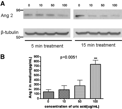
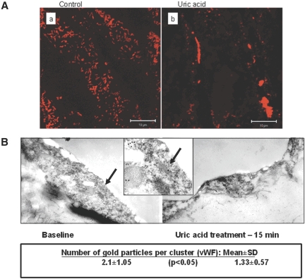
Recruitment of various stem and progenitor cells is crucial for the regeneration of an injured organ. Levels of uric acid, one of the prototypical "alarm signals," surge after ischemia-reperfusion injury. Exogenous uric acid rapidly mobilizes endothelial progenitor cells and hematopoietic stem cells and protects the kidney from ischemia. The relatively fast responses to uric acid suggest that preformed second messengers may be released from a storage pool. Here, it is reported that monosodium urate (MSU) results in exocytosis of Weibel-Palade bodies in vitro and in vivo, leading to the release of IL-8, von Willebrand factor, and angiopoietin 2 in the culture medium or circulation. Confocal and immunoelectron microscopy confirmed depletion of von Willebrand factor in MSU-treated aortic endothelial cells. Angiopoietin 2 alone induced exocytosis of Weibel-Palade bodies, mobilized hematopoietic stem cells and depleted splenic endothelial progenitor cells, partially reproducing the actions of MSU. In addition, pretreatment with angiopoietin 2 protected the kidneys from an ischemic insult, suggesting that the previously reported renoprotection conferred by MSU likely results from exocytosis of Weibel-Palade bodies. Furthermore, experiments with toll-like receptor 4 (TLR-4)-and TLR-2-deficient mice demonstrated that uric acid-induced exocytosis of Weibel-Palade bodies is mediated by TLR-4 and that uric acid-induced release of IL-8 requires both TLR-2 and TLR-4. In summary, these results suggest that exocytosis of Weibel-Palade bodies links postischemic repair with inflammation and mobilization of stem cells.


Figures

Figure 1.
Figure 1.
EPC and HSC in peripheral blood and spleen after MSU treatment. Mice received an injection of 50 μg/25 g MSU and were killed at indicated times. Blood and spleen were collected for isolation of EPC and HSC by FACS analysis, as detailed in the Concise Methods section (n = 5 in each group for each time-point). *P < 0.05.
Figure 2.
Figure 2.
Plasma levels of KC after monosodium urate injection. Here and below, * indicates P < 0.05, ** indicates P < 0.01, and *** indicates P < 0.001 unless otherwise indicated.
Figure 3.
Figure 3.
Exocytosis of WPB induced by UA. (A and B) Treatment of HUVEC with MSU (100 μg/ml) resulted in a rapid exocytosis of WPB and the loss of cellular vWF and angiopoietin 2. Images were obtained and processed as detailed in the Concise Methods section. Quantitative analysis of fluorescence intensity for A. Approximately 100 individual cells were analyzed in each of three independent experiments. (C and D) Dosage dependence (10, 50, and 100 μg/ml) of MSU effect on the expression of angiopoietin 2 and vWF. Quantitative analysis of fluorescence intensity. *Significant decrease in the expression of vWF and angiopoietin 2.
Figure 4.
Figure 4.
Decreased granularity (side scatter) of HUVEC after MSU treatment. (Right) Representative side-scatter tracings from control and MSU-treated HUVEC. Left) Summary of side-scatter experiments (P < 0.05, n = 3; Kolmogorov-Smirnov statistical analysis).
Figure 5.
Figure 5.
Time course and dosage-response of angiopoietin 2 expression. (A) Western blot analysis of HUVEC lysates. (B) ELISA detection of angiopoietin 2 in the conditioned medium. Note that the most prominent effect was detectable after 15 min of treatment with the reciprocal changes in the culture medium.
Figure 6.
Figure 6.
Decreased expression of angiopoietin 2 in the kidneys and a reciprocal increase in the serum level after MSU administration. (A, top) Typical results of Western blot analysis of angiopoietin 2 abundance in the mouse kidney 15 min after intraperitoneal injection of MSU (50 μg). (A, bottom) Summary of the results presented in A. (B) Serum angiopoietin 2 in mice after MSU treatment. *Significant difference from control (P = 0.046; n = 3).
Figure 7.
Figure 7.
Laser-scanning confocal microscopy of en face aorta and transmission immunoelectron microscopy of aortic endothelium. (A) Representative views of laser-scanning confocal microscopy of the endothelial layer of en face aorta from control and MSU-treated mice. (B) Immunoelectron microscopy of vWF in aortic endothelial cells. Note the labeling of vWF within WPB (insert) and reduction of immunodetectable vWF after MSU. vWF (10 nm gold) and angiopoietin 2 (6 nm gold particles) labeling of aortic endothelia at baseline (left) and 15 min after administration of MSU (right). Central panel provides a zoomed view (arrows) of WPB with vWF labeling. Angiopoietin 2 labeling is too sparse to be quantified. Quantification of vWF labeling showed a significant decline after UA. Magnification, ×29,000.
Figure 8.
Figure 8.
Effects of angiopoietin 2 on HSC and EPC. (A) Rapid elevation of HSC (CD150+/c-Kit+ cells) in peripheral blood and spleen 3 h after angiopoietin 2 injection. *P = 0.02. (B) Mobilization of the splenic pool of CD34+/Flk-1+ EPC 3 h after angiopoietin 2 injection. *P = 0.03 (n = 3 in each group for each time point).
Figure 9.
Figure 9.
Angiopoietin 2 pretreatment prevents renal functional deterioration after renal ischemia (n = 3 to 6; *P = 0.0005 control versus ischemia; P = 0.02 control versus ischemia+angiopoietin 2; P = 0.005 ischemia versus ischemia+angiopoietin 2).
Figure 10.
Figure 10.
Release of vWF and angiopoietin 2 in TLR-2 but not in TLR-4 knockout mice after administration of MSU. After administration of UA (50 μg/mouse), TLR-2–deficient mice (n = 8 at baseline, n = 5 at 30 min, and n = 3 at 3 h) within 30 min had increased plasma levels of vWF and angiopoietin 2. This effect was abrogated in TLR-4–deficient mice (n = 8 at baseline, n = 5 at 30 min, and n = 3 at 3 h). In contrast, plasma KC was not increased in both TLR-2–and TLR-4–deficient mice.
Figure 11.
Figure 11.
A schematic view of the mechanistic cascade triggered by renal ischemia-reperfusion acute and transient surge in UA, which acting on TLR-4 and TLR-2 leads to mobilization of different components of WPB and eventually determines the balance between the pro-repair and proinflammatory sequelae. Antagonistic relations between TLR-2 and TLR-4 may exist, but clarification of this will require further studies.

References

    1. Takahashi T, Kalka C, Masuda H, Chen D, Silver M, Kearney M, Magner M, Isner JM, Asahara T: Ischemia- and cytokine-induced mobilization of bone marrow-derived endothelial progenitor cells for neovascularization. Nat Med 5: 434–438, 1999 - PubMed
    1. Shintani S, Murohara T, Ikeda H, Ueno T, Honma T, Katoh A, Sasaki K, Shimada T, Oike Y, Imaizumi T: Mobilization of endothelial progenitor cells in patients with acute myocardial infarction. Circulation 103: 2776–2779, 2001 - PubMed
    1. Gill M, Dias S, Hattori K, Rivera ML, Hicklin D, Witte L, Girardi L, Yurt R, Himel H, Rafii S: Vascular trauma induces rapid but transient mobilization of VEGFR2(+)AC133(+) endothelial precursor cells. Circ Res 88: 167–174, 2001 - PubMed
    1. Patschan D, Krupincza K, Patschan S, Zhang Z, Hamby C, Goligorsky MS: Dynamics of mobilization and homing of endothelial progenitor cells after acute renal ischemia: Modulation by ischemic preconditioning. Am J Physiol Renal Physiol 291: F176–F185, 2006 - PubMed
    1. Zimmet JM, Hare JM: Nitroso-redox interactions in the cardiovascular system. Circulation 114: 1531–1544, 2006 - PubMed

Publication types