. 2008 Aug 26;105(34):12194-9.
doi: 10.1073/pnas.0803154105. Epub 2008 Aug 20.

Structural basis of Clostridium perfringens toxin complex formation

Affiliations

Structural basis of Clostridium perfringens toxin complex formation

Jarrett J Adams et al. Proc Natl Acad Sci U S A. .

Abstract

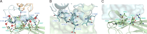
The virulent properties of the common human and livestock pathogen Clostridium perfringens are attributable to a formidable battery of toxins. Among these are a number of large and highly modular carbohydrate-active enzymes, including the mu-toxin and sialidases, whose catalytic properties are consistent with degradation of the mucosal layer of the human gut, glycosaminoglycans, and other cellular glycans found throughout the body. The conservation of noncatalytic ancillary modules among these enzymes suggests they make significant contributions to the overall functionality of the toxins. Here, we describe the structural basis of an ultra-tight interaction (K(a) = 1.44 x 10(11) M(-1)) between the X82 and dockerin modules, which are found throughout numerous C. perfringens carbohydrate-active enzymes. Extensive hydrogen-bonding and van der Waals contacts between the X82 and dockerin modules give rise to the observed high affinity. The mu-toxin dockerin module in this complex is positioned approximately 180 degrees relative to the orientation of the dockerin modules on the cohesin module surface within cellulolytic complexes. These observations represent a unique property of these clostridial toxins whereby they can associate into large, noncovalent multitoxin complexes that allow potentiation of the activities of the individual toxins by combining complementary toxin specificities.


Conflict of interest statement

The authors declare no conflict of interest.

Figures

Fig. 1.
Fig. 1.
Modular architecture of Clostridium perfringens X82- and Doc-containing glycoside hydrolases. The FIVAR, Doc, and X82 modules used in this study are shaded in orange, light blue, and green, respectively. GHXX, glycoside hydrolase catalytic domains (shaded gray) where XX represents the family number; CBM32, family 32 carbohydrate-binding module; UNK, modules having unknown function and little sequence identity with other protein modules; F, FIVAR modules; Doc, dockerin-like modules; CBM40, family 40 carbohydrate-binding module; X82, cohesin-like family 82 X-module; FN3, fibronectin III-like module; CBM50, family 50 carbohydrate-binding module; ConA-like, Con A-like module; BIG_2, group 2 bacterial Ig-like domain.
Fig. 2.
Fig. 2.
The ultrahigh affinity of the C. perfringens X82–Dockerin interaction. (A) ELISA-based binding specificities. X82 modules from family 3 (CpGH3), 20 (CpGH20), 31 (CpGH31), and 84 (CpGH84C) glycoside hydrolases and the “large” sialidase (NanJ) fused to a CBM were probed with Doc modules from the μ-toxin, and family 2 (CpGH2), 31 (CpGH31), and 95 (CpGH95) glycoside hydrolases, which were fused to a G. stearothermophilus xylanase T6. (+) and (−) indicate detectable and no detectable binding, respectively. (B) Isothermal titration calorimetric analysis of the CpGH84CX82-μ-toxin FIVAR-Doc interaction at 30°C. (Upper) Raw heat measurements. (Lower) Integrated heats after correction for heats of dilution as determined from the heats of ligand additions at the excess of saturation. The curves represent the best fit to a single-site model. (C) Temperature dependence of ΔH for the CpGH84CX82-μ-toxin FIVAR-Doc interaction. ΔH values were determined at 30°C, 35°C, and 37°C. (D) Differential scanning calorimetric denaturation profiles of 171 μM CpGH84CX82 (X82), 198 μM μ-toxin FIVAR-Doc (FivarDoc), and 18.2 μM CpGH84C-FIVAR-Doc complex. All samples contained 5 mM CaCl2.
Fig. 3.
Fig. 3.
Structure of a C. perfringens X82–Doc complex. Ribbon representation of the CpGH84CX82-μ-toxin FIVAR-Doc complex with CpGH84CX82 depicted in green, the FIVAR shown in orange, and the Doc module in light blue. The N- and C-termini are colored labeled accordingly. The calcium ions are depicted as purple spheres.
Fig. 4.
Fig. 4.
C. perfringens X82–Doc intermolecular contacts. (A) CpGH84CX82-μ-toxin Doc interface hydrogen-bonding network, with hydrogen-bond contacts shown as red dashed lines and water molecules as red spheres. (B) Ribbon representation of μ-toxin Doc, displaying residues involved in intermolecular van der Waals contacts as stick modules on the CpGH84CX82 surface. (C) Ribbon representation of CpGH84CX82, displaying residues involved in intermolecular van der Waals contacts as stick modules on the μ-toxin Doc surface. CpGH84CX82 and μ-toxin Doc are colored green and light blue, respectively. Residues are labeled in one-letter code, and numbered and colored accordingly.
Fig. 5.
Fig. 5.
Structural homology and interface residue conservation displayed by the C. perfringens X82 modules. (A) Backbone overlay (N, Cα, C′ atoms) of CpGH84CX82 (green) from the CpGH84CX82-μ-toxin FIVAR-Doc complex structure and isolated NanJX82 (salmon). (B) Overlay of the 9a-8-3-6-5 X82 faces and from CpGH84CX82 (green) and NanJX82 (salmon) depicting the Doc recognition residues of CpGH84CX82 and the analogous residues in NanJX82 as stick models. Residues are identified by one-letter code, and colored and numbered accordingly.
Fig. 6.
Fig. 6.
Variation of Doc orientations in clostridial complexes. Ribbon representations of (A) C. perfringens μ-toxin Doc (light blue) on CpGH84CX82 (light green); (B) native C. thermocellum type-I Doc (red) on type-I cohesin (yellow) (16); (C) C. thermocellum Ser45Ala/Thr46Ala type-I Doc mutant (red) on type-I cohesin (yellow) (36); (D) C. thermocellum type-II Doc (emerald green) on type-II cohesin (slate blue) (13). The helices within the two F-hand motifs of the Doc modules are identified as α1 and α2, respectively.

References

    1. Rood JI, Cole ST. Molecular genetics and pathogenesis of. Clostridium perfringens. Microbiol Rev. 1991;55:621–648. - PMC - PubMed
    1. Petit L, Gibert M, Popoff MR. Clostridium perfringens: Toxinotype and genotype. Trends Microbiol. 1999;7:104–110. - PubMed
    1. McDonel JL. Clostridium perfringens toxins (type A, B, C, D, E) Pharmacol Ther. 1980;10:617–655. - PubMed
    1. Myers GS, et al. Skewed genomic variability in strains of the toxigenic bacterial pathogen, Clostridium perfringens. Genome Res. 2006;16:1031–1040. - PMC - PubMed
    1. Shimizu T, et al. Complete genome sequence of. Clostridium perfringens, an anaerobic flesh-eater. Proc Natl Acad Sci USA. 2002;99:996–1001. - PMC - PubMed

Publication types