. 2008 Nov;19(11):4730-7.
doi: 10.1091/mbc.e08-06-0665. Epub 2008 Aug 20.

CLASP modulates microtubule-cortex interaction during self-organization of acentrosomal microtubules

Affiliations

CLASP modulates microtubule-cortex interaction during self-organization of acentrosomal microtubules

J Christian Ambrose et al. Mol Biol Cell. 2008 Nov.

Abstract

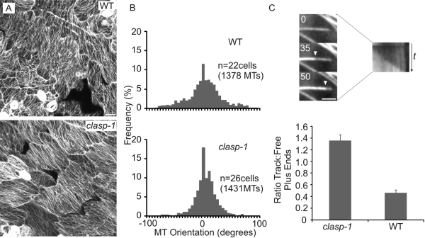
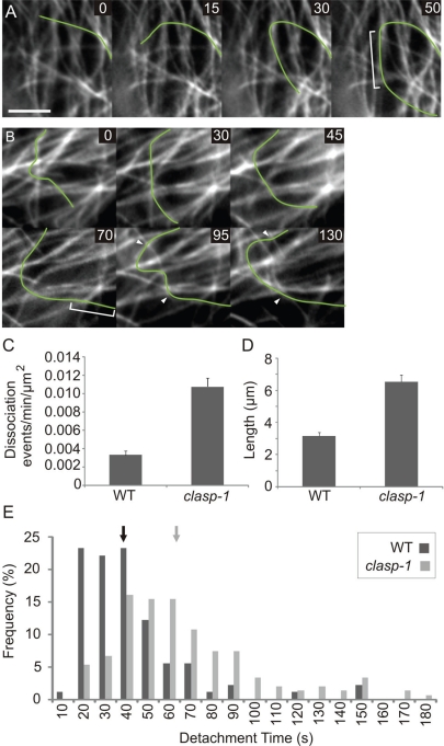
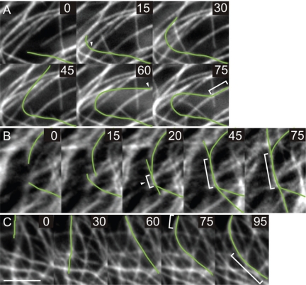
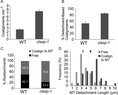
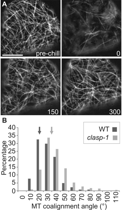
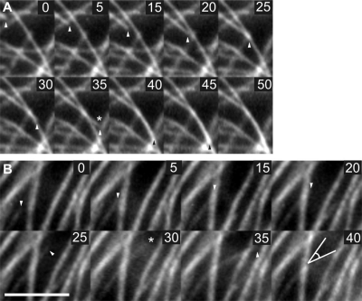
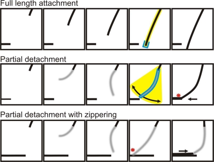
CLASP proteins associate with either the plus ends or sidewalls of microtubules depending on the subcellular location and cell type. In plant cells, CLASP's distribution along the full length of microtubules corresponds with the uniform anchorage of microtubules to the cell cortex. Using live cell imaging, we show here that loss of CLASP in Arabidopsis thaliana results in partial detachment of microtubules from the cortex. The detached portions undergo extensive waving, distortion, and changes in orientation, particularly when exposed to the forces of cytoplasmic streaming. These deviations from the normal linear polymerization trajectories increase the likelihood of intermicrotubule encounters that are favorable for subsequent bundle formation. Consistent with this, cortical microtubules in clasp-1 leaf epidermal cells are hyper-parallel. On the basis of these data, we identify a novel mechanism where modulation of CLASP activity governs microtubule-cortex attachment, thereby contributing to self-organization of cortical microtubules.


Figures

Figure 1.
Figure 1.
Cortical MTs in clasp-1 cotyledon epidermal cells are hyper-parallel. (A) Confocal images of cotyledon adaxial epidermal cells in wild type and clasp-1. Images are Z-projections of stacks taken at the lower middle region of the leaf. (B) Histogram of MT angular distributions in wild-type and clasp-1 cotyledon epidermal cells. Angles are normalized to each cell, where the 0° angle defines the predominant MT orientation. (C) A higher proportion of MT plus ends reside along other MTs in clasp-1 (n = 976) compared with wild type (n = 837). The image series shows an example of an MT growing along another “track” MT. A kymograph is shown at right. Data for C is taken from 15 cells each. Bar, (A) 20 μm, (C) 2.5 μm. Times in C are seconds.
Figure 2.
Figure 2.
Cortical MTs in clasp-1 cotyledon epidermal cells exhibit compromised cortex attachment. (A) Image sequence showing a typical MT detachment–reattachment event in clasp-1. On reassociation, the MT aligns with a cortex-bound MT (region of MT–MT overlap denoted by brackets). Corresponds to Supplementary Movie S4. (B) Very large MT detachment in clasp-1, accompanied by partial reattachment. Partial reattachment of the leading end occurs at 70 s (attached portion denoted by brackets). After partial reattachment, extensive MT distortions between the two points of cortical anchoring are apparent (arrowheads). Corresponds to Supplementary Movie S5. (C) Frequency of MT cortical detachments in wild-type and clasp-1 cotyledon epidermal cells. Detachment was defined by the MT moving out of the focal plane at some point during observation and/or any lateral swinging of the MT. (D) Mean length (μm) of MT detachments in wild-type and clasp-1 cotyledon epidermal cells. (E) MT detachment times (seconds) in wild-type and clasp-1 cotyledon epidermal cells. Means are denoted by arrows. Data are means ± SEM. Bar, 5 μm.
Figure 3.
Figure 3.
Cortical MT detachment behaviors often result in interactions between MTs in clasp-1. (A) Reattachment to a cortical MT upon reassociation. Arrowhead indicates the start of the detachment episode, which is coincident with encounter of the MT plus end with an obstacle MT. Brackets denote coalignment region between MTs after reattachment. Corresponds to Supplementary Movie S6. (B) Coalignment of two cortically detached MTs upon encounter. Note the gradual zippering and straightening of the region of overlap as it forms (brackets). Corresponds to Supplementary Movie S7. (C) MT bundle detachment. The bundle snaps into alignment with cortically associated MTs in two places (brackets). Corresponds to Supplementary Movie S8. Bar, 5 μm. Times are in seconds.
Figure 4.
Figure 4.
clasp-1 mutants exhibit enhanced interactions between MTs, and greater MT detachment is associated with an increased ability to bundle with other MTs. (A) Bundling events per minute in a 20 × 20-μm box in clasp-1 and wild type. (B) Percentage of MT bundle formations associated with a cortical detachment of one or both of the MTs. The proportion of detachment-associated bundling events is significantly higher in clasp-1. (C) Proportions of reattachments to cortex (free) or to another MT (bundle with MT) in clasp-1 and wild type. MTs reattach to other MTs more frequently in clasp-1. (D) Longer MT detachment lengths result in bundling with other MTs more frequently than short detachment lengths in wild type. Means of each class are indicated by arrows. Data are means ± SEM. All comparisons are statistically significant (p < 0.001).
Figure 5.
Figure 5.
Cells lacking CLASP allow more permissive MT coalignment. (A) An example of the cold-depolymerization assay used to allow unbiased quantification of MT coalignment. Shown is a wild-type cell before and after chilling for 4 min at −20°C. MTs recover rapidly over the 300 s observation period. Most MTs are depolymerized at the start of recovery, and by 300 s most MTs have returned. Corresponds to Supplementary Movie S9. (B) Frequency distributions of wild-type and clasp-1 MT coalignment angles in recovering cold-depolymerized cells. Arrows denote means that are significantly different (p < 0.001, t test). Bar, 10 μm.
Figure 6.
Figure 6.
Encounter of MT plus ends with obstacle MTs can result in detachment from the cortex. (A) Detachment resulting in coalignment with the obstacle MT. This sequence is from a wild-type cell. Asterisk denotes the brief detachment that occurs upon encounter with the obstacle MT. Coalignment occurs with the obstacle MT after this dissociation, at 40 s. (B) Detachment resulting in reorientation to a new growth trajectory upon reattachment. This sequence is from a clasp-1 cell. Bar, 10 μm.
Figure 7.
Figure 7.
Hypothetical model for organization of cortical MT arrays. Top panel, a strongly anchored MT, and middle panel, a partially detached MT (gray region). Yellow shading indicates the area of exploration, which is increased by the partial detachment. Blue outline denotes the surface area available for bundle formation. Red asterisk indicates initial contact points. Partial detachment allows exploration of a larger volume with a greater surface area, and the MT therefore captures and bundles with another MT. Bottom panel, an example of zippering from an initial contact point. Cortical detachment permits initial MT–MT contact points to propagate as MTs zipper together in a polymerization-independent manner. We propose that these processes occur in all cells, but are enhanced in the absence of CLASP. In this way, CLASP indirectly affects MT–MT interactions via maintaining strong association of MTs along their length with the cell cortex. The fact that MTs remain partially attached in the absence of CLASP indicates the presence of other cortical anchoring factors.

References

    1. Akhmanova A., et al. Clasps are CLIP-115 and -170 associating proteins involved in the regional regulation of microtubule dynamics in motile fibroblasts. Cell. 2001;104:923–935. - PubMed
    1. Ambrose J. C., Shoji T., Kotzer A. M., Pighin J. A., Wasteneys G. O. The Arabidopsis CLASP gene encodes a microtubule-associated protein involved in cell expansion and division. Plant Cell. 2007;19:2763–2775. - PMC - PubMed
    1. Bartolini F., Gundersen G. G. Generation of noncentrosomal microtubule arrays. J. Cell Sci. 2006;119:4155–4163. - PubMed
    1. Bratman S. V., Chang F. Stabilization of overlapping microtubules by fission yeast CLASP. Dev. Cell. 2007;13:812–827. - PMC - PubMed
    1. Chan J., Calder G. M., Doonan J. H., Lloyd C. W. EB1 reveals mobile microtubule nucleation sites in Arabidopsis. Nat. Cell Biol. 2003;5:967–971. - PubMed

Publication types

MeSH terms

Substances