

Prevalence of unsuspected pancreatic cysts on MDCT
- PMID: 18716113
- PMCID: PMC2692243
- DOI: 10.2214/AJR.07.3340
Prevalence of unsuspected pancreatic cysts on MDCT
Abstract
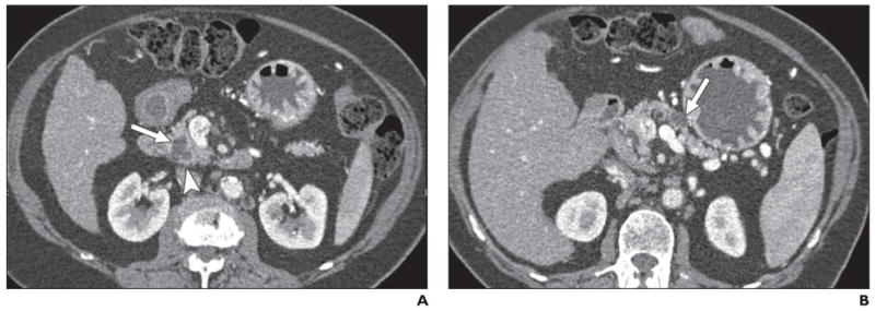
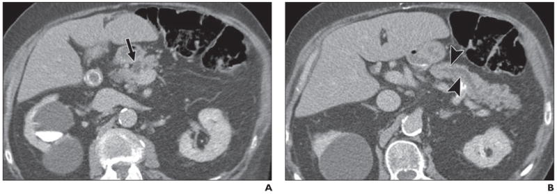
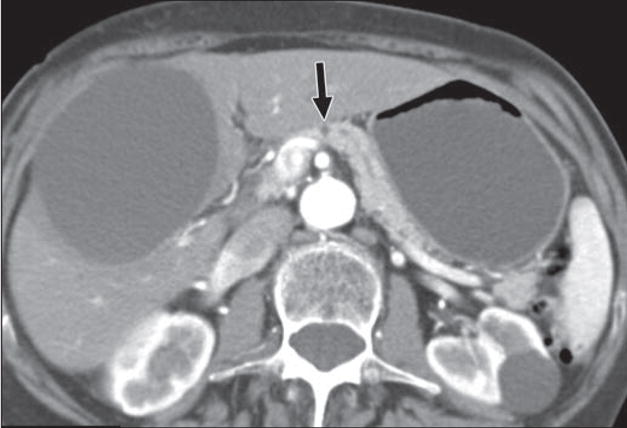
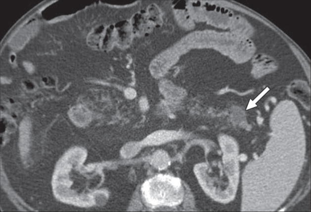
Objective: Current generation MDCT technology facilitates identification of small, nonenhancing lesions in the pancreas. The objective of this study was to determine the prevalence of findings of unsuspected pancreatic cysts on 16-MDCT in a population of adult outpatients imaged for disease unrelated to the pancreas.
Materials and methods: Contrast-enhanced MDCT scans of the abdomen were reviewed from 2,832 consecutive examinations to identify pancreatic cysts. Patients with a history of pancreatic lesions or predisposing factors for pancreatic disease or who were referred for pancreatic CT were excluded.
Results: A total of 73 patients had pancreatic cysts, representing a prevalence of 2.6 per 100 patients (95% CI, 2.0-3.2). Cysts ranged in size from 2 to 38 mm (mean, 8.9 mm) and were solitary in 85% of cases. Analysis of demographic information showed a strong correlation between pancreatic cysts and age, with no cysts identified among patients under 40 years and a prevalence of 8.7 per 100 (95% CI, 4.6-12.9) in individuals from 80 to 89 years. After controlling for age, cysts were more common in individuals of the Asian race than all other race categories, with an odds ratio of 3.57 (95% CI, 1.05-12.13). There was no difference by sex in the prevalence of cysts (p = 0.527); however, cysts were on average 3.6 mm larger (p = 0.014) in men than women.
Conclusion: In this outpatient population, the prevalence of unsuspected pancreatic cysts identified on 16-MDCT was 2.6%. Cyst presence strongly correlated with increasing age and the Asian race.
Figures
References
-
- American Cancer Society. Cancer, facts and figures 2007. [Accessed September 17, 2007]. www.cancer.org/downloads/STT/CAFF2007PWSecured.pdf.
-
- Winter JM, Cameron JL, Campbell KA, et al. 1423 pancreaticoduodenectomies for pancreatic cancer: a single-institution experience. J Gastrointest Surg. 2006;10:1199–1210. - PubMed
-
- Jemal A, Siegel R, Ward E, Murray T, Xu J, Thun MJ. Cancer statistics, 2007. CA Cancer J Clin. 2007;57:43–66. - PubMed
-
- Hruban RH, Pitman MB, Klimstra DS. Atlas of tumor pathology: tumors of the pancreas. 4. Washington, DC: American Registry of Pathology and Armed Forces Institute of Pathology; 2007.
-
- Hruban RH, Takaori K, Canto M, et al. Clinical importance of precursor lesions in the pancreas. J Hepatobiliary Pancreat Surg. 2007;14:255–263. - PubMed
Publication types
MeSH terms
Grants and funding
LinkOut - more resources
Full Text Sources
Other Literature Sources
Medical
