. 2008 Aug 20;28(34):8470-6.
doi: 10.1523/JNEUROSCI.0860-08.2008.

A fast mode of membrane fusion dependent on tight SNARE zippering

Affiliations

A fast mode of membrane fusion dependent on tight SNARE zippering

Marine Bretou et al. J Neurosci. .

Abstract

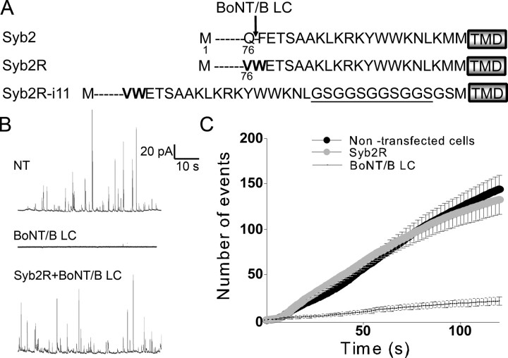
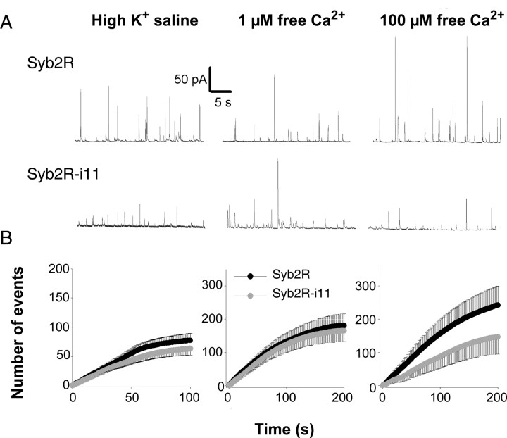
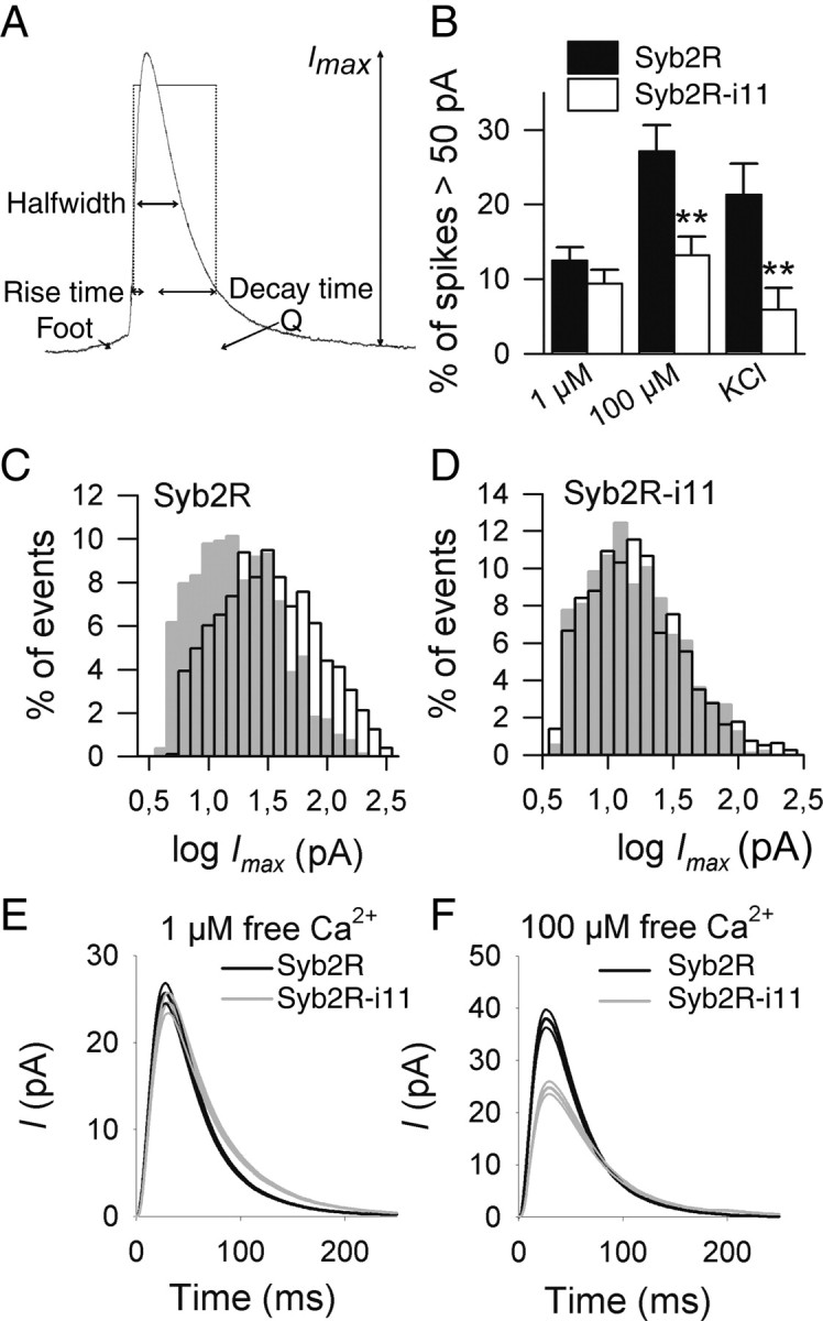
SNARE (soluble N-ethylmaleimide-sensitive factor attachment protein receptor) proteins have a key role in membrane fusion. It is commonly assumed that pairing of SNARE proteins anchored in opposing membranes overcomes the repulsion energy between membranes, thereby catalyzing fusion. In this study, we have increased the distance between the coiled-coil SNARE motif and the transmembrane domain of the vesicular SNARE synaptobrevin-2 by insertion of a flexible linker to analyze how an increased intermembrane distance affects exocytosis. Synaptobrevin-2 lengthening did not change the frequency of exocytotic events measured at 1 mum free calcium but prevented the increase in the secretory activity triggered by higher calcium concentration. Exocytotic events monitored in adrenal chromaffin cells by means of carbon fiber amperometry were classified in two groups according to the rate and extent of fusion pore expansion. Lengthening the juxtamembrane region of synaptobrevin-2 severely reduced the occurrence of rapid single events, leaving slow ones unchanged. It also impaired the increase in the fast-fusion mode that normally follows elevation of intracellular Ca2+ levels. We conclude that mild stimuli trigger slow fusion events that do not rely on a short intermembrane distance. In contrast, a short intermembrane distance mediated by tight zippering of SNAREs is essential to a component of the secretory response elicited by robust stimuli and characterized by rapid dilation of the fusion pore.


Figures

Figure 1.
Figure 1.
The secretory activity of chromaffin cells is abolished by BoNT/B light chain and restored by coexpression of Syb2R. A, Amino acid sequences of Syb2 and mutants. BoNT/B light chain cleaves Syb2 at Q76, as indicated. The transmembrane domain (TMD) is boxed. Point mutations that protect Syb2R from the proteolytic action of BoNT/B are in bold. Underlined amino acids were inserted to increase the distance between the SNARE motif and the transmembrane domain. Moreover, two amino acids were modified, K94G and M95S, to introduce the linkers (see Materials and Methods). The name of the construct (Syb2R-i11) refers to the number of inserted amino acids. B, Examples of amperometric recordings of a nontransfected chromaffin cell (top, NT), a chromaffin cell expressing BoNT/B light chain (middle, BoNT/B LC), and a chromaffin cell coexpressing Syb2R and BoNT/B (bottom, Syb2R+BoNT/B LC). C, Cumulated numbers of exocytic events (mean ± SE) as a function of time for nontransfected chromaffin cells (21 cells), cells expressing BoNT/B (20 cells), and cells coexpressing Syb2R and BoNT/B (50 cells).
Figure 2.
Figure 2.
Inserting a linker (i11) prevents the occurrence of high-amplitude spikes and slightly affects the overall frequency of exocytotic events. A, B, Chromaffin cells coexpressing BoNT/B and either Syb2R or Syb2R-i11 were stimulated by high K+ saline (left panels) or permeabilized with digitonin in the presence of 1 (middle panels) or 100 (right panels) μm free Ca2+, as indicated. A, Representative amperometric recordings of Syb2R-expressing (top) or Syb2R-i11-expressing (bottom) cells. B, Cumulated numbers of exocytic events (mean ± SE) as a function of time. Spikes were obtained from 23 (high K+), 40 (1 μm), or 28 (100 μm) cells.
Figure 3.
Figure 3.
High calcium concentration increases Imax values in Syb2R but not in Syb2R-i11-expressing cells. A, Representation of an amperometric spike. Different parameters are depicted: amplitude (Imax), quantal charge (Q), halfwidth, 20–90% rise time, and 90–20% decay time. B, Shown is the mean (±SE) percentage of spikes with an amplitude >50 pA recorded in control cells (Syb2R, filled bars) or in cells expressing Syb2R-i11 (open bars) on stimulation with high K+ or on digitonin permeabilization at either 1 or 100 μm free Ca2+. Spikes were monitored from 11 to 15 cells in each condition. **p < 0.01. C, D, Raising the free Ca2+ concentration from 1 μm (gray bars) to 100 μm (open bars) shifted the distribution of log Imax values to the right in Syb2R-expressing (C) but not in Syb2R-i11-expressing (D) cells. Distributions were computed from 821 (Syb2R, 1 μm), 1045 (Syb2R, 100 μm), 965 (Syb2R-i11, 1 μm), and 797 (Syb2R-i11, 100 μm) spikes. E, The “mean” amperometric spike from Syb2R (black curve)- or Syb2R-i11 (gray curve)-expressing cells at 1 μm free calcium. All events were aligned at their onset (spikes with foot were omitted) and averaged. Shown is the mean (±SE) of >420 events. F, Averaged spike at 100 μm calcium.
Figure 4.
Figure 4.
Syb2 lengthening reduces the proportion of rapid spikes. Chromaffin cells were transfected with Syb2R (A, D, G, J) or Syb2R-i11 (B, E, H, K) and stimulated by local perfusion of digitonin in the presence of 1 μm (A–C) or 100 μm (D–L) free Ca2+. A total of 821 (A, C) or 1045 (D–L) amperometric spikes from Syb2R-expressing cells and 965 (B, C) or 797 (D–L) spikes from Syb2R-i11-expressing cells were analyzed. A, D, Distribution of the values of the maximal spike rise velocity (smax) in control cells (Syb2R) at 1 μm (A) or 100 μm (D) free Ca2+. Frequency plots indicate the existence of two populations of spikes that can be well fitted with two Gaussian functions (black curves). The dotted line is the sum of the two normal density functions. Fitting with a single normal density function gives a lower r2 value (0.77). B, E, Distribution of smax values in cells expressing Syb2R-i11. This distribution is also better fitted by two normal density functions (gray curves) rather than by a single one (r2 = 0.76). C, F, Merge of the Gaussian fits indicating that the proportion of rapid spikes is higher in Syb2R- than in Syb2R-i11-expressing cells. G–I, Distribution of the values of log rise time is also bimodal in Syb2R (G)-expressing cells. This is less clear in Syb2R-i11 cells because the fit to a single Gaussian function is almost as good (r2 = 0.903). The proportion of spikes with a short rise time is strongly reduced in Syb2R-i11 cells compared with control ones (I). J–L, Frequency plots of the log-transformed values of 90–20% decay time. The distribution is clearly bimodal in Syb2R cells (J). In contrast, in Syb2R-i11 cells (K) it is well fitted with a single density function similar to the one that describes the slow spikes in Syb2R cells (L).

References

    1. Albillos A, Dernick G, Horstmann H, Almers W, Alvarez de Toledo G, Lindau M. The exocytotic event in chromaffin cells revealed by patch amperometry. Nature. 1997;389:509–512. - PubMed
    1. Amatore C, Bouret Y, Travis ER, Wightman RM. Interplay between membrane dynamics, diffusion and swelling pressure governs individual vesicular exocytotic events during release of adrenaline by chromaffin cells. Biochimie. 2000;82:481–496. - PubMed
    1. Chen Y, Xu Y, Zhang F, Shin YK. Constitutive versus regulated SNARE assembly: a structural basis. EMBO J. 2004;23:681–689. - PMC - PubMed
    1. Chernomordik LV, Zimmerberg J, Kozlov MM. Membranes of the world unite! J Cell Biol. 2006;175:201–207. - PMC - PubMed
    1. Chizmadzhev YA, Cohen FS, Shcherbakov A, Zimmerberg J. Membrane mechanics can account for fusion pore dilation in stages. Biophys J. 1995;69:2489–2500. - PMC - PubMed

Publication types

MeSH terms

Substances