

Regenerating articular tissue by converging technologies
- PMID: 18716660
- PMCID: PMC2515637
- DOI: 10.1371/journal.pone.0003032
Regenerating articular tissue by converging technologies
Abstract
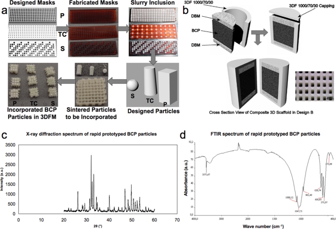
Scaffolds for osteochondral tissue engineering should provide mechanical stability, while offering specific signals for chondral and bone regeneration with a completely interconnected porous network for cell migration, attachment, and proliferation. Composites of polymers and ceramics are often considered to satisfy these requirements. As such methods largely rely on interfacial bonding between the ceramic and polymer phase, they may often compromise the use of the interface as an instrument to direct cell fate. Alternatively, here, we have designed hybrid 3D scaffolds using a novel concept based on biomaterial assembly, thereby omitting the drawbacks of interfacial bonding. Rapid prototyped ceramic particles were integrated into the pores of polymeric 3D fiber-deposited (3DF) matrices and infused with demineralized bone matrix (DBM) to obtain constructs that display the mechanical robustness of ceramics and the flexibility of polymers, mimicking bone tissue properties. Ostechondral scaffolds were then fabricated by directly depositing a 3DF structure optimized for cartilage regeneration adjacent to the bone scaffold. Stem cell seeded scaffolds regenerated both cartilage and bone in vivo.
Conflict of interest statement
Figures


Similar articles
-
Combining technologies to create bioactive hybrid scaffolds for bone tissue engineering.Biomatter. 2013 Apr-Jun;3(2):e23705. doi: 10.4161/biom.23705. Epub 2013 Jan 1. Biomatter. 2013. PMID: 23507924 Free PMC article.
-
Development of hybrid scaffolds using ceramic and hydrogel for articular cartilage tissue regeneration.J Biomed Mater Res A. 2015 Apr;103(4):1404-13. doi: 10.1002/jbm.a.35276. Epub 2014 Jul 28. J Biomed Mater Res A. 2015. PMID: 25044835
-
A composite scaffold of MSC affinity peptide-modified demineralized bone matrix particles and chitosan hydrogel for cartilage regeneration.Sci Rep. 2015 Dec 3;5:17802. doi: 10.1038/srep17802. Sci Rep. 2015. PMID: 26632447 Free PMC article.
-
Porous Scaffolds for Regeneration of Cartilage, Bone and Osteochondral Tissue.Adv Exp Med Biol. 2018;1058:171-191. doi: 10.1007/978-3-319-76711-6_8. Adv Exp Med Biol. 2018. PMID: 29691822 Review.
-
Unlike bone, cartilage regeneration remains elusive.Science. 2012 Nov 16;338(6109):917-21. doi: 10.1126/science.1222454. Science. 2012. PMID: 23161992 Free PMC article. Review.
Cited by
-
Combining regenerative medicine strategies to provide durable reconstructive options: auricular cartilage tissue engineering.Stem Cell Res Ther. 2016 Jan 28;7:19. doi: 10.1186/s13287-015-0273-0. Stem Cell Res Ther. 2016. PMID: 26822227 Free PMC article. Review.
-
Improvement of PHBV scaffolds with bioglass for cartilage tissue engineering.PLoS One. 2013 Aug 9;8(8):e71563. doi: 10.1371/journal.pone.0071563. eCollection 2013. PLoS One. 2013. PMID: 23951190 Free PMC article.
-
Combining technologies to create bioactive hybrid scaffolds for bone tissue engineering.Biomatter. 2013 Apr-Jun;3(2):e23705. doi: 10.4161/biom.23705. Epub 2013 Jan 1. Biomatter. 2013. PMID: 23507924 Free PMC article.
References
-
- Guldberg VM. Natural history of autografts and allografts. In: Older J, editor. Bone Implant Grafting. London: Springer; 1992. pp. 9–12.
-
- Hunzinker EB. Articular cartilage repair: basic science and clinical progress. A review of the current status and prospects. Osteoarthritis and Cartilage. 2001;10:432–463. - PubMed
-
- Athanasiou KA, Zhu C, Lanctot DR, Agrawal CM, Wang X. Fundamentals of biomechanics in tissue engineering of bone. Tissue Eng. 2000;6:361–381. - PubMed
-
- Laurencin CT, Ambrosio AM, Borden MD, Cooper JA., Jr Tissue engineering: orthopedic applications. Annu Rev Biomed Eng. 1999;1:19–46. - PubMed
-
- Martin I, Miot S, Barbero A, Jakob M, Wendt D. Osteochondral tissue engineering. J Biomech 2006 - PubMed
Publication types
MeSH terms
Substances
LinkOut - more resources
Full Text Sources
Other Literature Sources
Molecular Biology Databases
Research Materials
