. 2008 Sep 19;3(9):555-66.
doi: 10.1021/cb800059u. Epub 2008 Aug 22.

Chain dynamics of nascent polypeptides emerging from the ribosome

Affiliations

Chain dynamics of nascent polypeptides emerging from the ribosome

Jamie P Ellis et al. ACS Chem Biol. .

Abstract

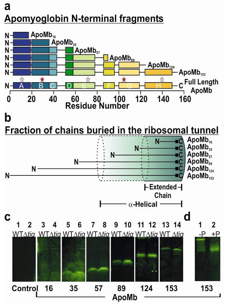
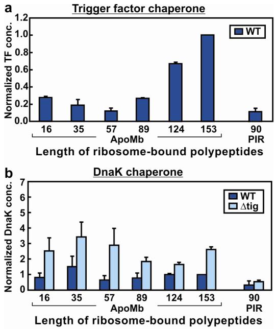
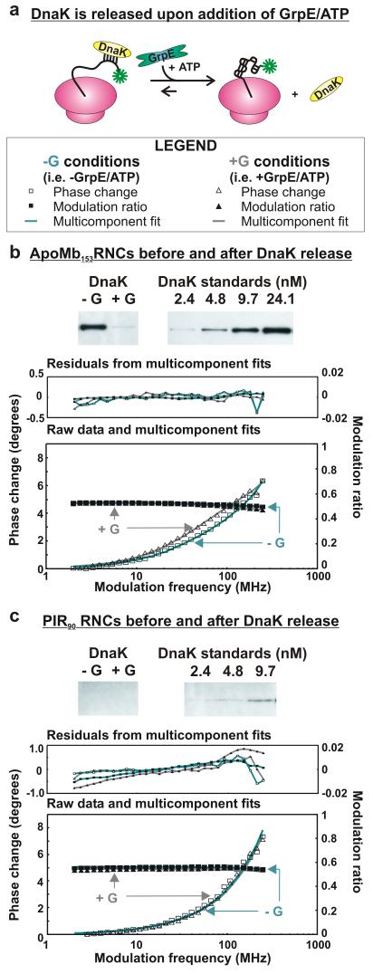
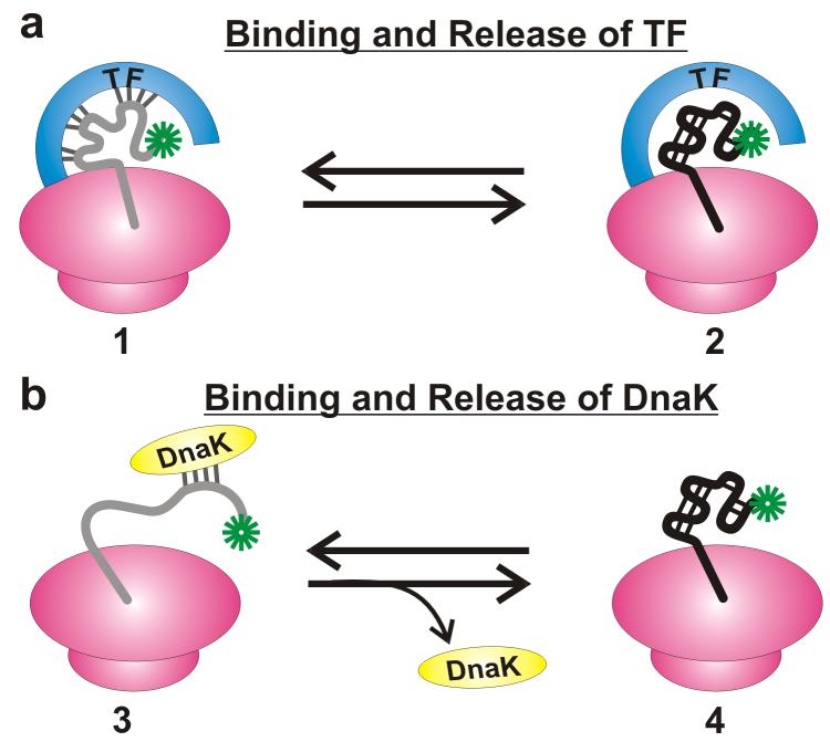
Very little is known about the conformation of polypeptides emerging from the ribosome during protein biosynthesis. Here, we explore the dynamics of ribosome-bound nascent polypeptides and proteins in Escherichia coli by dynamic fluorescence depolarization and assess the population of cotranslationally active chaperones trigger factor (TF) and DnaK. E. coli cell-free technology and fluorophore-linked E. coli Met-tRNA f Met enable selective site-specific labeling of nascent proteins at the N-terminal methionine. For the first time, direct spectroscopic evidence captures the generation of independent nascent chain motions for a single-domain protein emerging from the ribosome (apparent rotational correlation time approximately 5 ns), during the intermediate and late stages of polypeptide elongation. Such motions are detected only for a sequence encoding a globular protein and not for a natively unfolded control, suggesting that the independent nascent chain dynamics may be a signature of folding-competent sequences. In summary, we observe multicomponent, severely rotationally restricted, and strongly chain length/sequence-dependent nascent chain dynamics.


Figures

Figure 1
Figure 1
General features of ApoMb nascent chains. a) Schematic representation of apoMb ribosome-bound nascent polypeptides examined in this work. Chain lengths of N-terminal fragments are marked as subscripts. Colored segments (labeled A-H) denote α-helical regions of native full length apoMb. Stars indicate the experimentally detected high- (red) and low- (pink) affinity DnaK binding sites (48). b) Pictorial illustration of the expected fraction of nascent polypeptides buried inside the ribosomal exit tunnel (length ca. 100 Å, (1-3)) for limiting fully α-helical (1.5 Å /residue) and fully extended (3.5 Å /residue) conformations (see also Supporting Information). c) SDS-PAGE analysis of nascent apoMb polypeptides N-terminally tagged with BODIPY-FL-Met. Expression products for both wild-type (WT) and trigger factor-deficient (Δtig) cell-free systems are displayed. Samples in this panel are shown after treatment with puromycin (Puro). d) Verification of the fully ribosome-bound status of the nascent chains by treatment of ribosome-associated peptidyl-tRNAs with Puro. In the presence of the ribosome, Puro catalyzes the conversion of peptidyl-tRNA (lane 1) to peptidyl-Puro (lane 2), giving rise to a lower molecular weight species. As an example, the effect of Puro on apoMb153 RNCs is shown. Similar analysis was successfully performed on other nascent chains (Supporting Figure 1).
Figure 2
Figure 2
Normalized concentrations of TF and DnaK co-pelleting with RNCs. Normalized chaperone concentrations were obtained by dividing chaperone concentrations by ribosome-bound nascent chain levels (determined by fluorescence) and reporting the results relative to the values for the full length apoMb153 WT sample. Reported values are the average of two to six separate experiments. Uncertainties are reported as standard errors. a) Normalized concentrations of TF associating with translating ribosomes carrying nascent polypeptides. Cell-free expression was performed in WT cell-free systems. b) Normalized concentrations of DnaK associating with translating ribosomes carrying nascent polypeptides. Cell-free expression was performed in both WT and Δtig cell-free systems.
Figure 3
Figure 3
Technical aspects of dynamic fluorescence depolarization data collection. a) Representative physical model (3-component) for multiple independent molecular motions and matching equations applicable to rotational correlation times (τi) differing by at least one order of magnitude. The BODIPY-FL fluorophore is shown in green. Fi is the fractional amplitude for the anisotropy decay corresponding to each of the motions. In the case of 2-component RNC dynamics, the intermediate timescale (I) term is lost. b) Representative experimental fluorescence anisotropy data for short (apoMb16) and long (apoMb153) ribosome-bound nascent chains and associated 2- and 3-component fits, respectively. c) Multiexponential curve fitting (2- and 3-component analysis) and residuals for ribosome-bound ApoMb16. d) ApoMb153 analyzed similarly to panel c and 2-component fit with variable r0. e) Simulation illustrating the unique features of typical 2-and 3-component frequency-domain anisotropy decays. f) Expected phase changes upon varying the fraction of intermediate (fI) and fast timescale motions, to illustrate the progression from 2-(fI = 0) to 3-component decays. The 2- and 3-component phase change plots of panel e coincide with simulations A and C. g) Parameters used for the simulations in panel f.
Figure 4
Figure 4
Frequency domain dynamic fluorescence depolarization of ribosome-associated apoMb and PIR90 nascent chains generated in a wild-type E. coli cell-free system. ApoMb57 displays both 2- and 3-component dynamics. Data for a control sample of ribosome-released ApoMb153 are also shown. The cartoons on the far right highlight the proposed motions (in red) associated with each fluorescence phase. The BODIPY-FL fluorophore is shown in green.
Figure 5
Figure 5
Dynamic fluorescence depolarization of RNCs generated in a trigger factor-depleted (Δtig) cell-free system, before and after release of DnaK. a) Illustration of DnaK release induced by the addition of 2 μM GrpE, 30 μM ATP, and 500 μM KCl (+G conditions). b) Western blot showing that the concentration of apoMb153-RNC-bound DnaK is significantly reduced under +G conditions. This panel also shows the phase shift of the frequency domain fluorescence anisotropy of apoMb153-RNC upon release of DnaK. The shift corresponds to an enhancement in the fraction of ns motions. c) Western blot and fluorescence analysis (as in panel b) of PIR90. No shift from a 2-component fit is detected under +G conditions. Each experiment in panels b and c was repeated 3 times.
Figure 6
Figure 6
Proposed models for ribosome-bound nascent polypeptides consistent with the observed RNC dynamics. Both TF (a) and DnaK (b) are involved in binding/release events. The nascent chains are likely to interact with both chaperones, in the wild-type cell-free system. Only species of type 2 and 4 contribute to the 3–7 ns motion.
Figure 7
Figure 7
Simulations illustrating the expected domain sizes compatible with the experimentally detected rotational correlation time (4.5 ± 0.8 ns) of apoMb153 RNC motions. Three shapes were modeled: rigid fully extended polypeptide (prolate ellipsoid, black), semi-compact chain (prolate ellipsoid with fixed aspect ratio ρ=3.5, blue) and spherical collapsed chain (red). Correlation times were calculated (Supporting Information) assuming either the viscosity of water (a) or the viscosity sensed by ribosome-released full-length apoMb regarded as a globular and monomeric species in the resuspended RNC medium (b). Experimentally feasible chain lengths and structures fall under the shaded regions.

Comment in

References

    1. Schuwirth BS, Borovinskaya MA, et al. Structures of the bacterial ribosome at 3.5 .ANG. resolution. Science. 2005;310:827–834. - PubMed
    1. Nissen P, Hansen J, et al. The structural basis of ribosome activity in peptide bond synthesis. Science. 2000;289:920–930. - PubMed
    1. Harms J, Schluenzen F, et al. High Resolution Structure of the Large Ribosomal Subunit from a Mesophilic Eubacterium. Cell. 2001;107:679–688. - PubMed
    1. Baram D, Pyetan E, et al. Structure of trigger factor binding domain in biologically homologous complex with eubacterial ribosome reveals its chaperone action. Proc. Natl. Acad. Sci. U. S. A. 2005;102:12017–12022. - PMC - PubMed
    1. Ferbitz L, Maier T, et al. Trigger factor in complex with the ribosome forms a molecular cradle for nascent proteins. Nature. 2004;431:590–596. - PubMed

Publication types

MeSH terms