. 2009 Mar;11(3):589-600.
doi: 10.1089/ars.2008.2195.

Inhibition of ref-1 stimulates the production of reactive oxygen species and induces differentiation in adult cardiac stem cells

Affiliations

Inhibition of ref-1 stimulates the production of reactive oxygen species and induces differentiation in adult cardiac stem cells

Narasimman Gurusamy et al. Antioxid Redox Signal. 2009 Mar.

Abstract

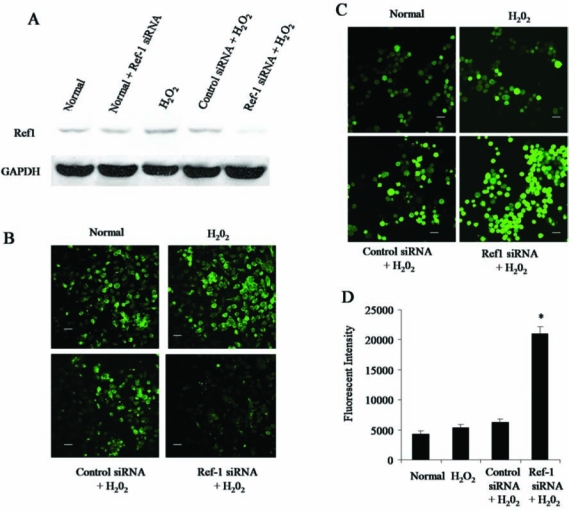
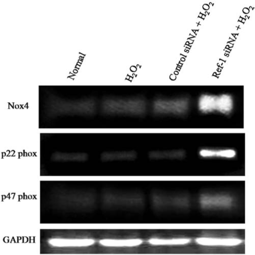
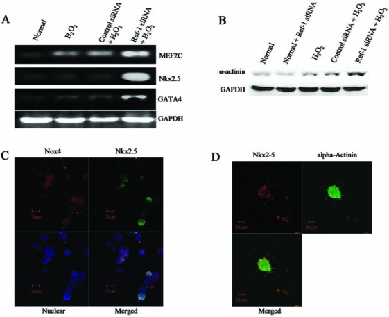
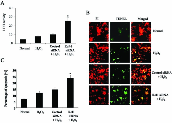
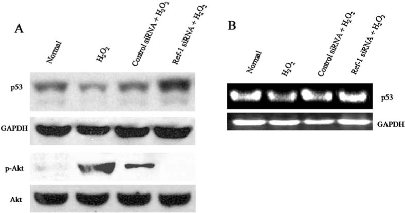
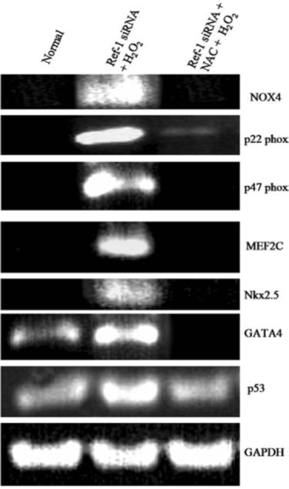
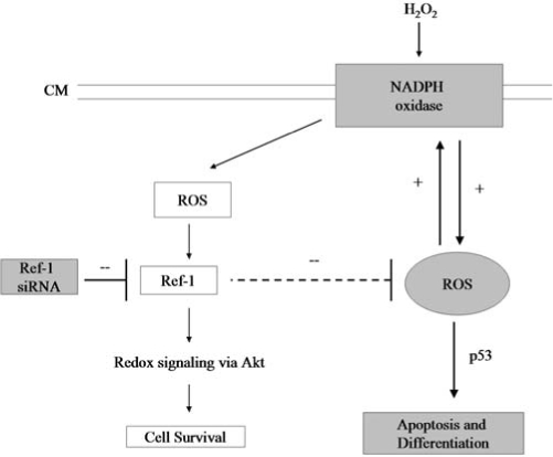
Redox effector protein-1 (Ref-1) plays an essential role in DNA repair and redox regulation of several transcription factors. In the present study, we examined the role of Ref-1 in maintaining the redox status and survivability of adult cardiac stem cells challenged with a subtoxic level of H2O2 under inhibition of Ref-1 by RNA interference. Treatment of cardiac stem cells with a low concentration of H2O2 induced Ref-1-mediated survival signaling through phosphorylation of Akt. However, Ref-1 inhibition followed by H2O2 treatment extensively induced the level of intracellular reactive oxygen species (ROS) through activation of the components of NADPH oxidase, like p22( phox ), p47( phox ), and Nox4. Cardiac differentiation markers (Nkx2.5, MEF2C, and GATA4), and cell death by apoptosis were significantly elevated in Ref-1 siRNA followed by H2O2-treated stem cells. Further, inhibition of Ref-1 increased the level of p53 but decreased the phosphorylation of Akt, a molecule involved in survival signaling. Treatment with ROS scavenger N-acetyl-L-cysteine attenuated Ref-1 siRNA-mediated activation of NADPH oxidase and cardiac differentiation. Taken together, these results indicate that Ref-1 plays an important role in maintaining the redox status of cardiac stem cells and protects them from oxidative injury-mediated cell death and differentiation.


Figures

FIG. 1.
FIG. 1.
Inhibition of Ref-1 increases the level of intracellular reactive oxygen species (ROS). Adult cardiac stem cells were treated with either control or Ref-1 siRNA followed by H2O2 (10 μM), as mentioned in Methods section. (A) Representative Western immunoblot showing the expression Ref-1 in the total cell lysate. GAPDH was used as loading control. (B) Representative confocal microscopic images showing the staining of Ref-1 in cells. (C) Representative confocal microscopic images showing the staining of cells with a cell-permeable redox-sensitive dye (CM-H2DCFDA) indicating the level of intracellular ROS. (D) Quantification of the average fluorescent intensity of cells stained with CM-H2DCFDA. Scale bar represents 20 μm in B and C. (For interpretation of the references to color in this figure legend, the reader is referred to the web version of this article at www.liebertonline.com/ars).
FIG. 2.
FIG. 2.
Ref-1 inhibition activates NADPH oxidase components. Adult cardiac stem cells were treated with either control or Ref-1 siRNA followed by H2O2 (10 μM), as mentioned in Methods. At the end of experimentation, cells were lysed in TRIzol reagent, and RNA was isolated. RT-PCR analysis was performed with specific primers against NOX4, p22phox, p47phox, and GAPDH.
FIG. 3.
FIG. 3.
Inhibition of Ref-1 induces differentiation in cardiac stem cells. Adult cardiac stem cells were treated with either control or Ref-1 siRNA followed by H2O2 (10 μM), as mentioned in Methods. (A) RNA was isolated from cells by using TRIzol reagent, and RT-PCR analysis was performed with specific primers against cardiac differentiation markers such as MEF2C, Nkx2.5, and GATA4. GAPDH was used as loading control. (B) Western immunoblotting was performed with total cell lysate by using specific antibodies against α-actinin. GAPDH was used as loading control. (C) Confocal microscopic images showing the Ref-1 inhibition–driven coexpression of NOX4 (red channel, Alexa Fluor 594) and Nkx2.5 (green channel, Alexa Fluor 488) in the nuclear (blue channel, To-Pro 1 iodide) region of the cell. (D) Confocal microscopic images showing the Ref-1 inhibition–mediated coexpression of Nkx2.5 (red channel) and α-actinin (green channel) in a differentiated cell. (For interpretation of the references to color in this figure legend, the reader is referred to the web version of this article at www.liebertonline.com/ars).
FIG. 4.
FIG. 4.
Ref-1 inhibition induces cell death. Adult cardiac stem cells were treated with either control or Ref-1 siRNA followed by H2O2 (10 μM), as mentioned in Methods. (A) The release of lactate dehydrogenase (LDH) enzyme from the cells was measured by using the spent culture medium obtained at the end of experimental period. (B) Fluorescent microscopic images showing the TUNEL staining of apoptotic cells (green channel) and nucleus (red channel, propidium iodide). (C) Quantification of apoptotic cells stained with TUNEL. The results are expressed in percentages. PI, propidium iodide. (For interpretation of the references to color in this figure legend, the reader is referred to the web version of this article at www.liebertonline.com/ars).
FIG. 5.
FIG. 5.
The regulation of p53 under Ref-1 siRNA treatment. Adult cardiac stem cells were treated with either control or Ref-1 siRNA followed by H2O2 (10 μM), as mentioned in Methods. (A) Representative Western immunoblot showing the expression of p53, GAPDH, p-Akt, and Akt in the total cell lysate. (B) RT-PCR analysis showing the expression of p53 and GAPDH.
FIG. 6.
FIG. 6.
Treatment with ROS scavenger attenuated Ref-1 siRNA–mediated cardiac differentiation. Adult cardiac stem cells were treated with either control or Ref-1 siRNA followed by H2O2 (10 μM), as mentioned in Methods. At the end of experimentation, cells were lysed in TRIzol reagent, and RNA was isolated. RT-PCR analysis was performed with specific primers against NOX4, p22phox, p47phox, MEF2C, Nkx2.5, GATA4, p53, and GAPDH.
FIG. 7.
FIG. 7.
Role of Ref-1 in the redox control of cardiac stem cells. When adult cardiac stem cells are treated with 10 μM H2O2, Ref-1–mediated redox signaling is activated, leading to the protection of cells from injury. However, inhibition of Ref-1 through Ref-1 siRNA treatment resulted in the production of massive amounts of reactive oxygen species (ROS) and activation of NADPH oxidase, leading to p53-mediated apoptosis and differentiation in cardiac stem cells. Discontinuous line, Ref-1 may inhibit the massive production of ROS. Inhibition of Ref-1 with Ref-1 siRNA induces the production of massive amounts of ROS, leading to apoptosis and differentiation in adult cardiac stem cells.

References

    1. Allen RG. Venkatraj VS. Oxidants and antioxidants in development and differentiation. J Nutr. 1992;122:631–635. - PubMed
    1. Almog N. Rotter V. Involvement of p53 in cell differentiation and development. Biochim Biophys Acta. 1997;1333:F1–F27. - PubMed
    1. Angkeow P. Deshpande SS. Qi B. Liu YX. Park YC. Jeon BH. Ozaki M. Irani K. Redox factor-1: an extra-nuclear role in the regulation of endothelial oxidative stress and apoptosis. Cell Death Differ. 2002;9:717–725. - PubMed
    1. Bachelder RE. Ribick MJ. Marchetti A. Falcioni R. Soddu S. Davis KR. Mercurio AM. p53 inhibits alpha 6 beta 4 integrin survival signaling by promoting the caspase 3-dependent cleavage of AKT/PKB. J Cell Biol. 1999;147:1063–1072. - PMC - PubMed
    1. Beckman BS. Balin AK. Allen RG. Superoxide dismutase induces differentiation of Friend erythroleukemia cells. J Cell Physiol. 1989;139:370–376. - PubMed

Publication types

Substances