. 2008 Aug 21:3:11.
doi: 10.1186/1750-1326-3-11.

Prion protein lacks robust cytoprotective activity in cultured cells

Affiliations

Prion protein lacks robust cytoprotective activity in cultured cells

Heather M Christensen et al. Mol Neurodegener. .

Abstract

Background: The physiological function of the cellular prion protein (PrPC) remains unknown. However, PrPC has been reported to possess a cytoprotective activity that prevents death of neurons and other cells after a toxic stimulus. To explore this effect further, we attempted to reproduce several of the assays in which a protective activity of PrP had been previously demonstrated in mammalian cells.

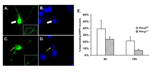
Results: In the first set of experiments, we found that PrP over-expression had a minimal effect on the death of MCF-7 breast carcinoma cells treated with TNF-alpha and Prn-p0/0 immortalized hippocampal neurons (HpL3-4 cells) subjected to serum deprivation. In the second set of assays, we observed only a small difference in viability between cerebellar granule neurons cultured from PrP-null and control mice in response to activation of endogenous or exogenous Bax.

Conclusion: Taken together, our results suggest either that cytoprotection is not a physiologically relevant activity of PrPC, or that PrPC-dependent protective pathways operative in vivo are not adequately modeled by these cell culture systems. We suggest that cell systems capable of mimicking the neurotoxic effects produced in transgenic mice by N-terminally deleted forms of PrP or Doppel may represent more useful tools for analyzing the cytoprotective function of PrPC.


Figures

Figure 1
Figure 1
Untransfected MCF-7 cells undergo cell death following treatment with recombinant TNF-α. (A) MCF-7 cells were treated with 100 ng/ml TNF-α, and cell viability was assessed using the MTT assay. Absorbance values are expressed as a percentage of those in non-treated control samples at each timepoint. Error bars are derived from triplicate wells of one representative experiment. (B) Flow cytometry analysis of untreated or TNF-α-treated MCF-7 cells. Cells were trypsinized, fixed in ethanol, and incubated with propidium iodide prior to analysis by flow cytometry. 10,000 cells were counted for each condition. Data are expressed as the percentage of cells containing sub-2n DNA content in one representative experiment.
Figure 2
Figure 2
Expression of human PrP mildly suppresses TNF-α-mediated death of MCF-7 breast carcinoma cells. (A) PrP expression was assessed by western blotting of three pools of vector-transfected MCF-7 cells (lanes 1–3) and six pools of MCF-7 cells transfected with a human PrP plasmid (lanes 4–9). (B) Pools of MCF-7 cells were treated with 100 ng/ml of recombinant TNF-α for 43 h. Cell viability was determined by the MTT assay. Absorbance values are expressed as a percentage of those in non-treated control samples. Data represent the mean ± SEM from 3 vector-transfected pools and 6 PrP-transfected pools. *p = 0.0042, human PrP vs. vector. (C) Pools of MCF-7 cells were treated with 100 ng/ml of recombinant TNF-α for 43 h. Cell viability was determined by flow cytometry after staining with propidium iodide. 10,000 cells were counted for each condition, and data are expressed as the percentage of cells containing sub-2n DNA. Data are derived from 4 MCF-7(hPrP) pools (mean ± SEM) and one vector control pool.
Figure 3
Figure 3
Stable expression of PrP weakly suppresses death of immortalized Prn-p0/0 hippocampal neurons after serum deprivation. (A) Two untransfected lines of Prn-p0/0 HpL cells (3–2 and 3–4) were subjected to 72 hours of serum deprivation. Cell viability was determined by MTT assay. Absorbance values are expressed as a percentage of those in non-treated control samples at each timepoint. Data are from one representative experiment. (B) Immunofluorescence analysis of mouse PrP expression in two PrP-expressing lines of HpL3-4 cells (4-1 and 4-2), and one vector-transfected line. PrP was detected by staining with the PrP antibody (green). Cells were counterstained with giantin, a marker for the Golgi apparatus (red). PrP expression was detectable on the cell surface and in the Golgi apparatus. Bar = 50 μm. (C) HpL3-4 cell lines were subjected to 96 hours of serum deprivation, after which cell viability was determined by flow cytometry after propidium iodide staining. 10,000 cells were counted for each condition. Data are expressed as the percentage of cells containing sub-2n DNA content, and represent the mean ± SEM from three independent experiments. *p < 0.01, PrP vs. vector. Relative PrP expression for each cell line is indicated below each set of bars by the number of "+" symbols.
Figure 4
Figure 4
PrP does not significantly reduce death of cerebellar granule neurons induced by exogenous Bax. (A-D) Representative fluorescence images of cerebellar granule neurons from Prn-p0/0 mice transfected with plasmids encoding EGFP and mouse Bax. Four days after transfection, cultures were stained with DAPI, and EGFP-positive neurons were scored as healthy (A, B) or apoptotic (C, D) based on morphological criteria. These criteria included the distribution of EGFP in the soma and neurites, and the appearance of DAPI staining in the nucleus (see text). (E) CGNs cultured from Prn-p+/+ or Prn-p0/0 pups were transfected with plasmids encoding Bax and EGFP. Cultures were fixed and stained with DAPI either 8 or 16 hours after transfection, and scored for apoptotic morphology. Data represent the mean ± SEM from at least three independent experiments. The difference between Prn-p0/0 and Prn-p+/+ neurons did not reach statistical significance at either time point (p > 0.05).
Figure 5
Figure 5
CGNs expressing PrP are slightly more resistant to cell death induced by potassium/serum deprivation. CGNs cultured from Prn-p0/0 and Prn-p+/+ mice were cultured in K25+S medium for 7 days, after which they were transferred to K5-S medium for the designated times. Cell viability was assessed by measurement of calcein fluorescence on a microplate fluorimeter. Fluorescence values in K5-S medium are expressed as a percentage of those in K25+5 medium at each time point. Data represent the mean ± SEM for at least two independent experiments. * p < 0.05, Prn-p+/+ vs. Prn-p0/0.

References

    1. Prusiner SB, (Ed) Prion Biology and Diseases. Second. Cold Spring Harbor, New York: Cold Spring Harbor Laboratory Press; 2004.
    1. Prusiner SB. Prions. Proc Natl Acad Sci USA. 1998;95:13363–13383. doi: 10.1073/pnas.95.23.13363. - DOI - PMC - PubMed
    1. Büeler H, Fischer M, Lang Y, Fluethmann H, Lipp H-P, DeArmond SJ, Prusiner SB, Aguet M, Weissmann C. Normal development and behavior of mice lacking the neuronal cell-surface PrP protein. Nature. 1992;356:577–582. doi: 10.1038/356577a0. - DOI - PubMed
    1. Manson JC, Clarke AR, Hooper ML, Aitchison L, McConnell I, Hope J. 129/Ola mice carrying a null mutation in PrP that abolishes mRNA production are developmentally normal. Mol Neurobiol. 1994;8:121–127. doi: 10.1007/BF02780662. - DOI - PubMed
    1. Mallucci GR, Ratte S, Asante EA, Linehan J, Gowland I, Jefferys JG, Collinge J. Post-natal knockout of prion protein alters hippocampal CA1 properties, but does not result in neurodegeneration. EMBO J. 2002;21:202–210. doi: 10.1093/emboj/21.3.202. - DOI - PMC - PubMed