. 2008 Oct;244(1-2):45-50.
doi: 10.1016/j.heares.2008.07.009. Epub 2008 Aug 6.

Ontogeny of serotonin and serotonin2A receptors in rat auditory cortex

Affiliations

Ontogeny of serotonin and serotonin2A receptors in rat auditory cortex

Gregory J Basura et al. Hear Res. 2008 Oct.

Abstract

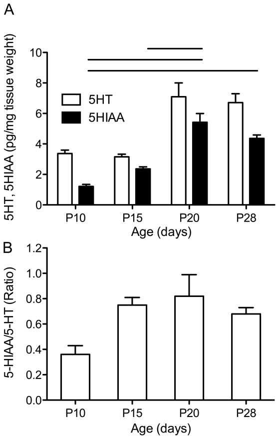
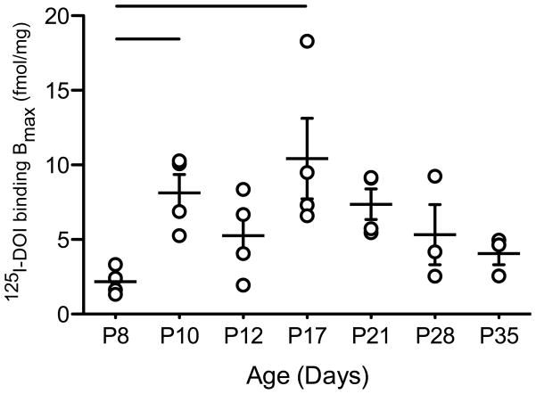
Maturation of the mammalian cerebral cortex is, in part, dependent upon multiple coordinated afferent neurotransmitter systems and receptor-mediated cellular linkages during early postnatal development. Given that serotonin (5-HT) is one such system, the present study was designed to specifically evaluate 5-HT tissue content as well as 5-HT(2A) receptor protein levels within the developing auditory cortex (AC). Using high performance liquid chromatography (HPLC), 5-HT and the metabolite, 5-hydroxyindoleacetic acid (5-HIAA), was measured in isolated AC, which demonstrated a developmental dynamic, reaching young adult levels early during the second week of postnatal development. Radioligand binding of 5-HT(2A) receptors with the 5-HT(2A/2C) receptor agonist, (125)I-DOI ((+/-)-1-(2,5-dimethoxy-4-iodophenyl)-2-aminopropane HCl; in the presence of SB206553, a selective 5-HT(2C) receptor antagonist, also demonstrated a developmental trend, whereby receptor protein levels reached young adult levels at the end of the first postnatal week (P8), significantly increased at P10 and at P17, and decreased back to levels not significantly different from P8 thereafter. Immunocytochemical labeling of 5-HT(2A) receptors and confocal microscopy revealed that 5-HT(2A) receptors are largely localized on layer II/III pyramidal cell bodies and apical dendrites within AC. When considered together, the results of the present study suggest that 5-HT, likely through 5-HT(2A) receptors, may play an important role in early postnatal AC development.


Figures

Figure 1
Figure 1
5-HT and 5-HIAA tissue levels in developing AC. A. Relative levels of 5-HT and 5-HIAA expressed as picograms of monoamine per mg of tissue weight in normal postnatal AC development. Despite a significant effect of age, no individual 5-HT age comparisons were significantly different. ANOVA followed by a Tukey’s multiple comparison test shows that 5-HIAA levels were significantly different at P10 vs P20 (p<0.01); P10 vs P28 (p<0.05) and P15 vs P20 (p<0.05). B. 5-HT/5-HIAA ratio indicative of 5-HT metabolism in AC during normal development. Young adult (P28) ratio is reached by P15. The only significantly different levels are found between P10 and P20 (p<0.05). The lines above the bar graph link significantly different comparisons.
Figure 2
Figure 2
A. Whole brain autoradiograph images demonstrating 125I-DOI labeling of 5-HT2A receptors within the rat AC during postnatal development from P8 to P35. Dark bars represent the limits of AC (including Te1 and Te3) and the regional of analysis. B. Right: Enlarged image of P35 rat brain demonstrating area of analysis (bars) representing AC used for quantification in each section. Note the robust signal dorsal to the rhinal fissure (arrow) extending from AC to other cortical areas. Left: Higher magnification shows that the 5-HT2A receptor binding in AC is highest in layers II and III, and lower in other layers. This anatomic location was confirmed with immunocytochemistry and confocal microscopy (see Figure 4).
Figure 3
Figure 3
Quantified 125I-DOI radio-ligand binding of 5-HT2A receptors within the developing AC expressed as fmol/mg. The graph represents pixel densities, converted to tissue binding levels, from radio-ligand binding of 5- HT2A receptors from P8 to P35 (autoradiographs shown in Fig. 2). Data are shown as the individual measurements from each animal (open circles), and the mean and SEM for each age. Statistical analysis was performed using a non-parametric one-way ANOVA (Kruskal-Wallis, significance at p<0.05). 5-HT2A receptor expression increases to young adult levels within the first two weeks of postnatal development, and declines thereafter. Post-hoc tests (Dunn’s) showed that significant increases were seen between P8 and P10 and P8 and P17. There was no difference between P8 and measurements at P21, P28 and P35, suggesting a significant decline following P17. The line above the bar graph links significantly different comparisons.
Figure 4
Figure 4
5-HT2A receptor expression in AC using immunocytochemistry and confocal imaging. A. Low power image (10X objective) showing the position of the rhinal fissure (arrow) and dorsally located 5-HT2A receptor immunoreactive dendrites within the AC using a 5-HT2A receptor monoclonal antibody (PharMingen; 1:500; note: bars outline AC region containing Te1 and Te3 seen at higher magnification in panels B and C). B. High magnification (40X objective) from panel A showing 5-HT2A receptor immunoreactivity within apical dendrites of pyramidal neurons located within layers II/III of the AC. C. Negative control section in which the primary 5- HT2A receptor antibody was omitted from the assay. Note background staining but no labelling of cell bodies or fibers.

References

    1. Araneda R, Andrade R. 5-Hydroxytryptamine2 and 5-hydroxytryptamine 1A receptors mediate opposing responses on membrane excitability in rat association cortex. Neuroscience. 1991;40:399–412. - PubMed
    1. Azmitia EC, Segal M. An autoradiographic analysis of the differential ascending projections of the dorsal and median raphe nuclei in the rat. J Comp Neurol. 1978;179:641–667. - PubMed
    1. Basura GJ, Walker PD. Serotonin 2A receptor mRNA levels in the neonatal dopamine-depleted rat striatum remain upregulated following suppression of serotonin hyperinnervation. Brain Res Dev Brain Res. 1999;116:111–117. - PubMed
    1. Beique JC, Campbell B, Perring P, Hamblin MW, Walker P, Mladenovic L, Andrade R. Serotonergic regulation of membrane potential in developing rat prefrontal cortex: coordinated expression of 5-hydroxytryptamine (5-HT)1A, 5-HT2A, and 5-HT7 receptors. J Neurosci. 2004;24:4807–4817. - PMC - PubMed
    1. Bennett-Clarke CA, Chiaia NL, Rhoades RW. Thalamocortical afferents in rat transiently express high-affinity serotonin uptake sites. Brain Res. 1996b;733:301–306. - PubMed

Publication types