. 2008 Dec 31;164(3):300-11.
doi: 10.1016/j.resp.2008.07.019. Epub 2008 Jul 31.

Interleukin (IL)-1 regulates ozone-enhanced tracheal smooth muscle responsiveness by increasing substance P (SP) production in intrinsic airway neurons of ferret

Affiliations

Interleukin (IL)-1 regulates ozone-enhanced tracheal smooth muscle responsiveness by increasing substance P (SP) production in intrinsic airway neurons of ferret

Z-X Wu et al. Respir Physiol Neurobiol. .

Abstract

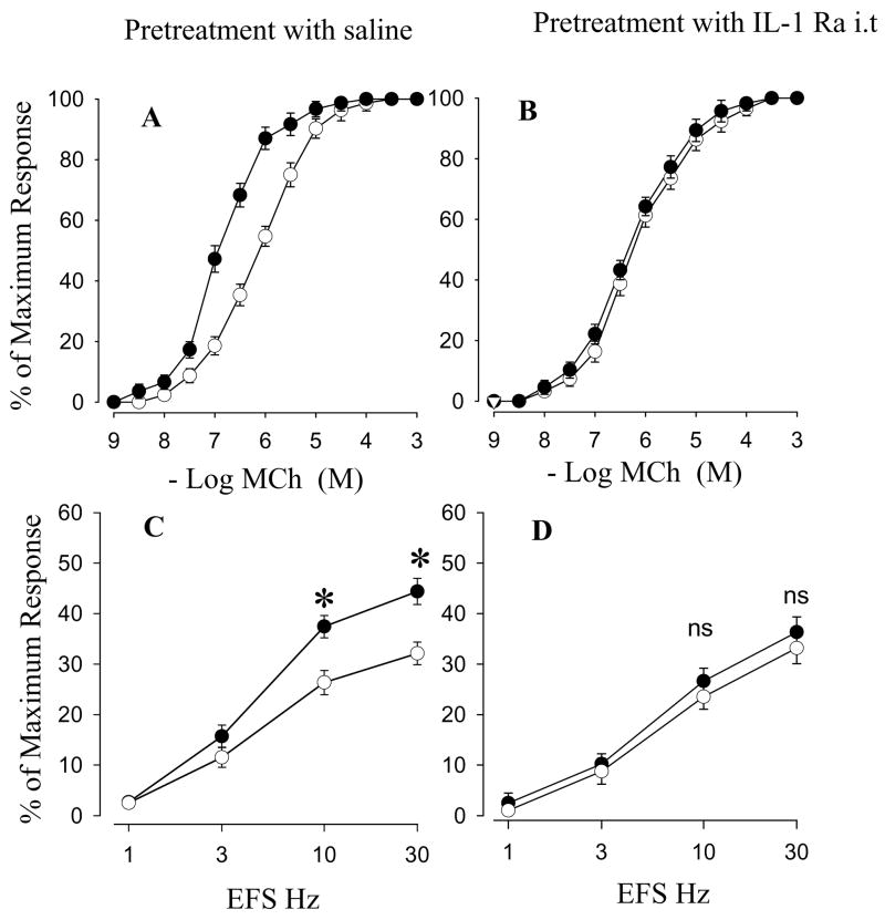
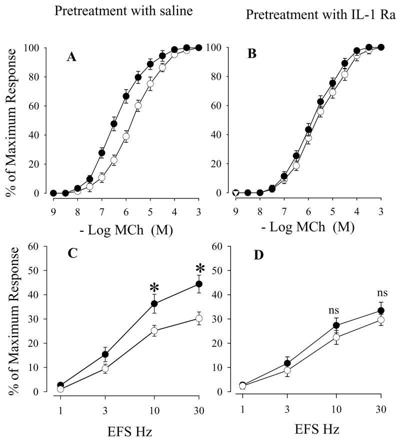
Exposure to ozone induces airway hyperresponsiveness (AHR) mediated partly by substance P (SP) released from nerve terminals of intrinsic airway neurons. Our recent studies showed that interleukin (IL)-1, an important multifunctional proinflammatory cytokine, increases synthesis and release of SP from intrinsic airway neurons. The purpose of this study is to investigate the possible involvement of endogenous IL-1 in modulating neural responses associated with ozone-enhanced airway responsiveness. Ferrets were exposed to 2ppm ozone or filtered air for 3h. IL-1 in the bronchoalveolar lavage (BAL) fluid was significantly increased in ozone-exposed animals and responses of tracheal smooth muscle to methacholine (MCh) and electrical field stimulation (EFS) were elevated significantly. Both the SP nerve fiber density in tracheal smooth muscle and the number of SP-containing neurons in airway ganglia were significantly increased following ozone exposure. Pretreatment with IL-1 receptor antagonist (IL-1 Ra) significantly diminished ozone-enhanced airway responses to EFS as well as ozone-increased SP in the airway. To selectively investigate intrinsic airway neurons, segments of ferret trachea were maintained in culture conditions for 24h to eliminate extrinsic contributions from sensory nerves. The segments were then exposed to 2ppm ozone in vitro for 3h. The changes of ozone-induced airway responses to MCh and EFS, and the SP levels in airway neurons paralleled those observed with in vivo ozone exposure. The ozone-enhanced airway responses and neuronal SP levels were inhibited by pretreatment with IL-1 Ra. These findings show that IL-1 is released during ozone exposure enhances airway responsiveness by modulating SP expression in airway neurons.


Figures

Figure 1
Figure 1
Effect of ozone exposure on IL-1 release in bronchoalveolar lavage fluid obtained from air and ozone exposed animals. Values are means ± SE; n = 5 in each group. IL-1 was measured by ELISA. * Significant difference between in vivo air and ozone exposed animals, P ≤0.05.
Figure 2
Figure 2
Effects of saline (A and C) or IL-1 Ra i.v (B and D) on cumulative concentration-response curves for MCh (A and B) and frequency-response curves for EFS (C and D) in tracheal smooth muscle after in vivo exposure to air (○) or ozone (●). Values are means ± SE; n = 5. The difference in MCh cumulative concentration-response curves between air and ozone exposure are presented in Table 2. *Significant difference in EFS between air and ozone exposure, P ≤ 0.05.
Figure 3
Figure 3
Effects of saline (A and C) or IL-1 Ra i.t (B and D) on cumulative concentration-response curves for MCh (A and B) and frequency-response curves for EFS (C and D) in tracheal smooth muscle after in vivo exposure to air (○) or ozone (●). Values are means ± SE; n = 5. The difference in MCh cumulative concentration-response curves between air and ozone exposure are presented in Table 2. *Significant difference in EFS between air and ozone exposure, P ≤0.05.
Figure 4
Figure 4
Effects of the different concentrations of IL-1 Ra on frequency-response curves for EFS in organotypic cultured tracheal smooth muscle prior to IL-1 treatment. Figure 4A shows that 10 ng/ml IL-1 enhanced airway smooth muscle responses to EFS and 200 ng/ml IL-1 Ra totally inhibited IL-1-enhanced airway smooth muscle responses to EFS. Figure 4B shows that effects of the different concentrations of IL-1 Ra on IL-1-enhanced airway smooth muscle responses to EFS at 10 Hz. Figure 4C shows the effects of the different concentrations of IL-1 Ra on IL-1-enhanced airway smooth muscle responses to EFS at 30 Hz. Values are means ± SE; * Significant difference in EFS between IL-1 with IL-1 Ra 0 ng/ml treatment and control (no IL-1 and no IL-1 Ra treatment) or IL-1 with IL-1 Ra 200 ng/ml treatment, P ≤0.05.
Figure 5
Figure 5
Effects of saline (A and C) or IL-1 receptor antagonist (B and D) on cumulative concentration-response curves for MCh (A and B) and frequency-response curves for EFS (C and D) in organotypic cultured tracheal smooth muscle after in vitro exposure to air (○) or ozone (●). Values are means ± SE; n = 5. The difference in MCh cumulative concentration-response curves between air and ozone in vitro exposure are presented in Table 3 *Significant difference in EFS between in vitro air and ozone exposure, P ≤0.05.
Figure 6
Figure 6
Fluorescence photomicrographs of substance P (SP)-immunoreactive nerve cell bodies within superficial muscular plexus and SP-immunoreactive nerve fiber density within tracheal smooth muscle in IL-1 Ra or saline-treated tracheal segments after in vitro exposure to air or ozone. A (air exposure with IL-1 Ra pretreatment): few SP-immunoreactive nerve fibers are present in tracheal smooth muscle (NFD of this micrograph is 0.20). B (ozone exposure with saline pretreatment): increased SP-immunoreactive nerve fibers in tracheal smooth muscle (NFD of this micrograph is 0.41). C (ozone exposure with IL-1 Ra pretreatment) decreased SP-immunoreactive nerve fibers in tracheal smooth muscle (NFD of this micrograph is 0.21). D (air exposure with IL-1 Ra pretreatment): negative SP-immunoreactive neurons are present in the SMP. E (ozone exposure with saline pretreatment): SP-immunoreactive cell bodies are increased in the SMP. F (ozone exposure with IL-1 Ra pretreatment): negative SP-immunoreactive neurons are seen. The images of the SP-containing nerve cell bodies in SMP and SP NFD in air exposure group with saline pretreatment (not shown) are the same as air exposure with IL-1 pretreatment. Magnification: x285.
Figure 7
Figure 7
Effects of IL-1 receptor antagonist treatment on SP-containing nerve cell bodies in SMP and LT and SP nerve fiber density in tracheal smooth muscle after in vitro exposure to air or ozone. Values are means ± SE; n = 5. *Significant difference between saline/ozone and other groups, P ≤0.05.
Figure 8
Figure 8
Effects of IL-1 receptor antagonist i.v on SP-containing nerve cell bodies in SMP and LT and SP nerve fiber density in tracheal smooth muscle after in vivo exposure to air or ozone. Values are means ± SE; n = 5. *Significant difference between saline/ozone and other groups, P ≤0.05.

References

    1. Arsalane K, Gosset P, Vanhee D, Voisin C, Hamid Q, Tonnel AB, Wallaert B. Ozone stimulates synthesis of inflammatory cytokines by alveolar macrophages in vitro. Am J Respir Cell Mol Biol. 1995;13:60–68. - PubMed
    1. Baker DG, McDonald DM, Basbaum CB, Mitchell RA. The architecture of nerves and ganglia of the ferret trachea as revealed by acetylcholinesterase histochemistry. J Comp Neurol. 1986;246:513–526. - PubMed
    1. Barchasz E, Naline E, Molimard M, Moreau J, Georges O, Emonds-Alt X, Advenier C. Interleukin-1beta-induced hyperresponsiveness to [Sar9,Met(O2)11]substance P in isolated human bronchi. Eur J Pharmacol. 1999;379:87–95. - PubMed
    1. Barnes PJ, Baraniuk JN, Belvisi MG. Neuropeptides in the respiratory tract. Am Rev Respir Dis. 1991;144:1391–1399. - PubMed
    1. Becker S, Clapp WA, Quay J, Frees KL, Koren HS, Schwartz DA. Compartmentalization of the inflammatory response to inhaled grain dust. Am J Respir Crit Care Med. 1999;160:1309–1318. - PubMed

Publication types

MeSH terms