. 2008 Aug 26;18(16):1256-61.
doi: 10.1016/j.cub.2008.07.092.

Evidence for an upper limit to mitotic spindle length

Affiliations

Evidence for an upper limit to mitotic spindle length

Martin Wühr et al. Curr Biol. .

Abstract

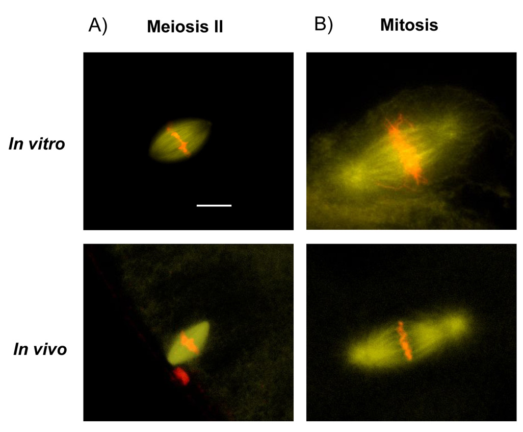
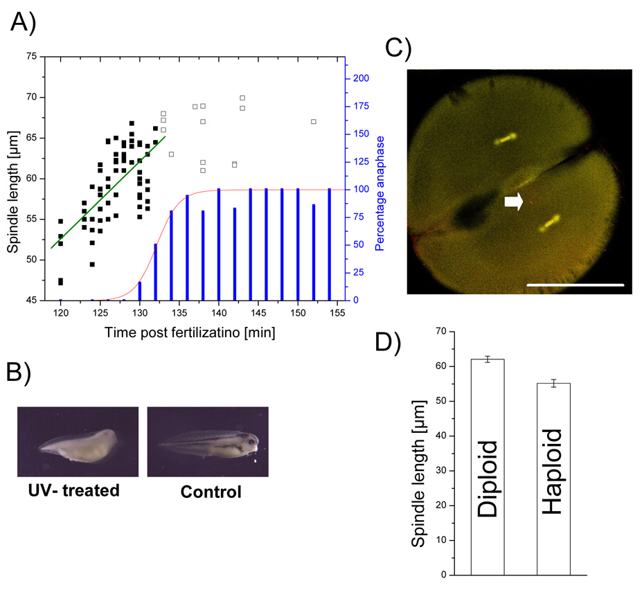
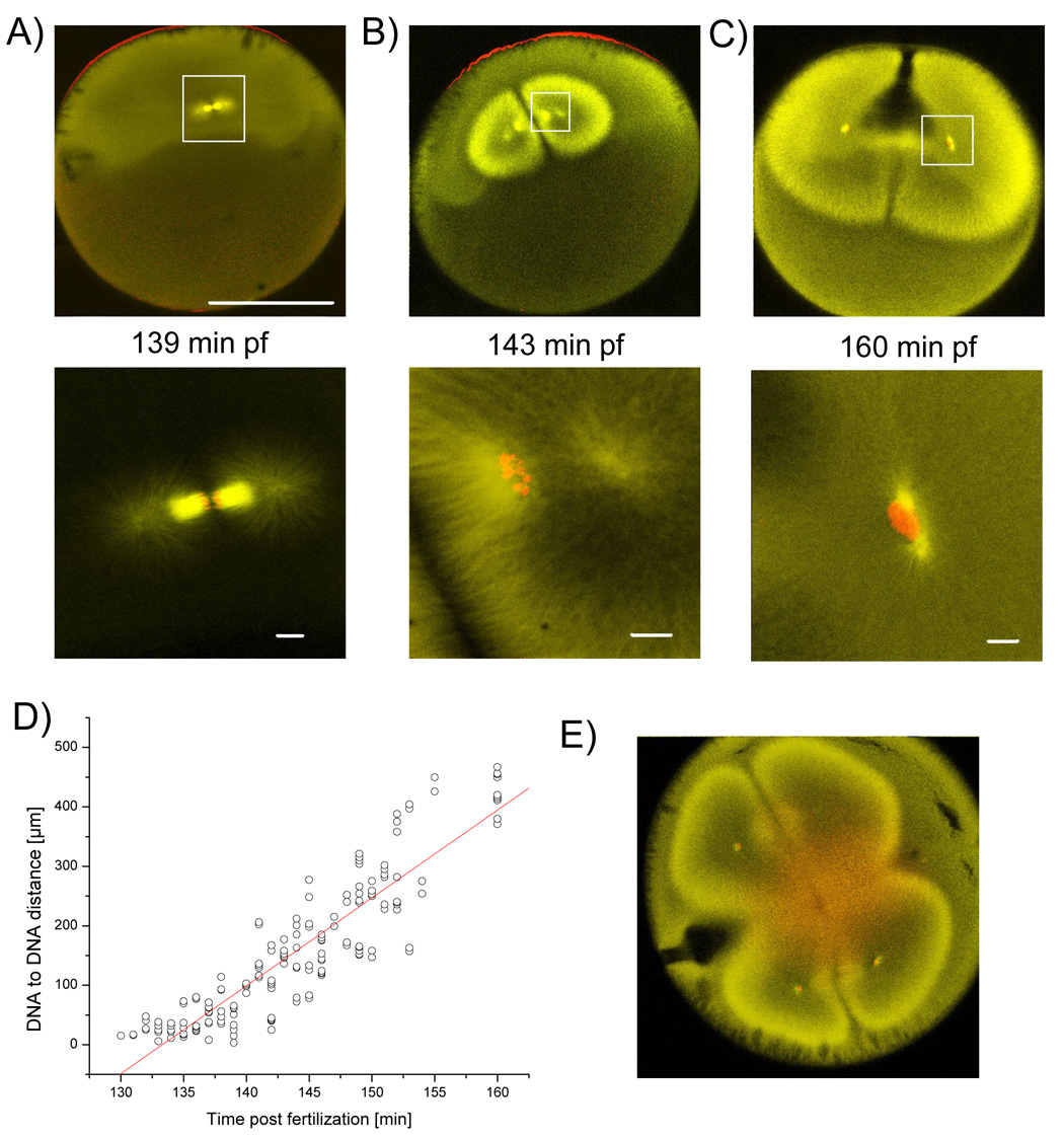
Size specification of macromolecular assemblies in the cytoplasm is poorly understood [1]. In principle, assemblies could scale with cell size or use intrinsic mechanisms. For the mitotic spindle, scaling with cell size is expected, because the function of this assembly is to physically move sister chromatids into the center of nascent daughter cells. Eggs of Xenopus laevis are among the largest cells known that cleave completely during cell division. Cell length in this organism changes by two orders of magnitude ( approximately 1200 microm to approximately 12 microm) while it develops from a fertilized egg into a tadpole [2]. We wondered whether, and how, mitotic spindle length and morphology adapt to function at these different length scales. Here, we show that spindle length increases with cell length in small cells, but in very large cells spindle length approaches an upper limit of approximately 60 microm. Further evidence for an upper limit to spindle length comes from an embryonic extract system that recapitulates mitotic spindle assembly in a test tube. We conclude that early mitotic spindle length in Xenopus laevis is uncoupled from cell length, reaching an upper bound determined by mechanisms that are intrinsic to the spindle.


Figures

Figure 1
Figure 1. Spindle size is uncoupled from cell size during first mitoses
X. laevis embryos at various stages of development were fixed and stained for tubulin (yellow) and DNA (red). A) Embryo at stage 8: animal pole with smaller cells and smaller spindles on top, vegetal pole with larger cells and larger spindles on bottom. B) Embryo at mitosis 7 animal part. C) Second mitotic spindle. White lines define spindle and cell size used throughout this paper. D) Egg arrested at metaphase of meiosis II with arrow pointing at spindle. Bar for upper row = 500 µm. Bar for lower row = 20 µm. E) Plot of cell size versus spindle size at different stages of development. Spindle size increases with cell size but asymptotically reaches an upper limit ~60 µm. Plot on the right is a zoom-in of smaller cells and spindles.
Figure 2
Figure 2. Embryonic extract is able to assemble mitotic spindles
DNA is shown in red and tubulin in yellow. A) Extract prepared from meiosis II arrested eggs assembles spindles that show similar morphology to meiotic in vivo spindles. B) Spindles in extract prepared from embryos were arrested in mitosis with addition of the APC-inhibitor EmiI. The spindles formed show similar morphology to mitotic in vivo spindles. Bar = 20 µm.
Figure 3
Figure 3. Halving the DNA content reduces spindle length by ~10%
A) Percentage of embryos (synchronously fertilized) in anaphase (blue bars) was fitted to a cumulative Gaussian distribution (red line), calculating the time for metaphase-anaphase transition at 132 ± 3 min (SD). Spindle length before peak of anaphase onset (full squares) was fitted linearly (green line) revealing spindle growth of 1.0 µm/min. Delayed spindles (shown as empty squares) were ignored for growth measurement as this would have systematically underestimated growth rate. B) Albino eggs were fertilized with UV-treated sperm from a pigmented male resulting in tadpoles with no pigments but haploid phenotype. Control developed with pigments and diploid phenotype C) Sperm derived DNA (arrow) is separate from spindles at two-cell stage of haploid embryo. Bar = 500 µm. D) Spindle mean length for haploids is 55.2 µm and therefore ~10 % shorter than diploids with 62.1 µm. Standard errors are 0.9 µm and 1.1 µm, respectively, with a statistically significant p-value of 0.005 %.
Figure 4
Figure 4. Relatively small spindle is compensated by enormous anaphase B like movement
Embryos of a synchronously fertilized population were fixed between first and second cytokineses and stained for tubulin (yellow) and DNA (red). A) At anaphase the astral microtubules start to elongate. B) Up to a DNA to DNA distance of ~180 µm, DNA is still condensed and surrounded by high staining of microtubules. Astral microtubules form a hollow structure. C) Nuclear envelope has reformed and finally the nuclei have been separated by ~400 µm, astral microtubules reach the cell cortex and cytokinesis starts. A–C) Bar for upper row = 500 µm, Bars in lower row = 20 µm. D) Plot of DNA to DNA distance versus time. Linear fit estimates speed of DNA separation at ~15 µm/min. E) Cytokinesis, but not separation of DNA, is inhibited by addition of 33 µg/ml of actin depolymerazing Cytochalasin B.

References

    1. Marshall WF. Cellular length control systems. Annu Rev Cell Dev Biol. 2004;20:677–693. - PubMed
    1. Montorzi M, Burgos MH, Falchuk KH. Xenopus laevis embryo development: arrest of epidermal cell differentiation by the chelating agent 1,10-phenanthroline. Mol Reprod Dev. 2000;55:75–82. - PubMed
    1. Gard DL, Cha BJ, Schroeder MM. Confocal immunofluorescence microscopy of microtubules, microtubule-associated proteins, and microtubule-organizing centers during amphibian oogenesis and early development. Curr Top Dev Biol. 1995;31:383–431. - PubMed
    1. Andersen SS, Ashford AJ, Tournebize R, Gavet O, Sobel A, Hyman AA, Karsenti E. Mitotic chromatin regulates phosphorylation of Stathmin/Op18. Nature. 1997;389:640–643. - PubMed
    1. Burbank KS, Mitchison TJ, Fisher DS. Slide-and-cluster models for spindle assembly. Curr Biol. 2007;17:1373–1383. - PubMed

Publication types

LinkOut - more resources